Volume 13, Issue 3 (10-2025)
Jorjani Biomed J 2025, 13(3): 10-16 |
Back to browse issues page


Download citation:
BibTeX | RIS | EndNote | Medlars | ProCite | Reference Manager | RefWorks
Send citation to:


BibTeX | RIS | EndNote | Medlars | ProCite | Reference Manager | RefWorks
Send citation to:
Nameni F, Jebraiili E. The effect of high-intensity interval training on cardiac angiogenesis and hypoxia-response in diabetic rats. Jorjani Biomed J 2025; 13 (3) :10-16
URL: http://goums.ac.ir/jorjanijournal/article-1-1071-en.html
URL: http://goums.ac.ir/jorjanijournal/article-1-1071-en.html


1- Department of Exercise Physiology, VaP.C., Islamic Azad University, Varamin Iran , namenifarah@gmail.com
2- Department of. Exercise Physiology, SR.C, Islamic Azad University, Tehran Iran
2- Department of. Exercise Physiology, SR.C, Islamic Azad University, Tehran Iran
Keywords: Vascular Endothelial Growth Factor A, HIF-1alpha protein, Human, High-Intensity Interval Training, Diabetes Mellitus
Full-Text [PDF 511 kb]
(3158 Downloads)
| Abstract (HTML) (11782 Views)
In the second week, when the rats were well familiarized with the pool, they were trained to the interval format by being taken out of the water several times after one minute of swimming using a rest plate and then placed back in the water. Forty-eight hours after the last familiarization session, the rats in the training group first performed a 5-minute warm-up exercise and then the main HIIT swimming workout, which consisted of 14 repetitions of 20-second swimming exercises with 10 seconds of rest between each repetition. At the end of each training session, a 5-minute cool-down was applied, and then all rats were dried and returned to their cages. This exercise program was performed for eight weeks (Three days per week, on alternate days). In the load-interval training, the weight applied in the first week was 7% of the body weight of each rat and was increased by 1% each week so that in the final (Eighth) week the rats swam with a weight equivalent to 14% of their body weight tied to the root of their tails. Training was performed in the evening. During the intervention period, the control group did not perform any training program (13). For the load, a small weighted bead attached to the tail by a band (Tail band) was used. Before each session, the weight was adjusted to the body weight of each rat using a scale. Water depth was at least twice the body length of the rats, and its temperature was controlled at a constant 30 ± 0.5 °C throughout all sessions by a digital thermometer. Under supervision, the rats were allowed to swim at their natural speed; the researcher simply monitored them and did not force or stimulate increased speed. Animal fatigue was assessed by observing swimming behavior, stopping, diving, or attempting to escape; additional rest was given in case of severe fatigue. During the first one to two weeks, the rats adapted to the protocol and were able to complete all repetitions without stopping.
Research protocol
To induce diabetes, streptozotocin (STZ; 60 mg/kg; Sigma-Aldrich, USA; ≥98% purity by HPLC) dissolved in 0.1 M citrate buffer (pH 4.5) was injected subcutaneously between the two ears. Seven days after injection, blood glucose was measured, and rats with blood glucose levels higher than 250 mg/dL were selected as diabetic samples. Blood glucose was measured before STZ injection and on days 0, 7, 14, 21, and 35. Ketamine and xylazine were used to anesthetize the animals at doses of 80 mg/kg and 10 mg/kg, respectively, administered intraperitoneally (i.p.). This dose produced deep and stable anesthesia for surgical interventions and cardiac biopsy. After sampling, hearts were removed and stored at −80 °C until RNA extraction. To examine the expression of vascular VEGF-A and hypoxia-inducible factor genes, total cellular RNA was extracted using an RNA extraction column kit (FavorPrep™ Tissue Total RNA Mini Kit) according to the manufacturer's instructions.
Sampling was performed 48 hours after the last training session and after an overnight fast. First, the rats were anesthetized, the chest was opened, and blood was taken directly from the heart to minimize trauma. Heart muscle was separated under sterile conditions. Blood samples taken directly from the heart were collected and stored in Falcon tubes.
Real-time PCR protocol
RNA extraction
Total RNA was extracted from heart tissue samples (Approximately 50 mg per sample) stored at -80 °C using a commercial RNA isolation kit (RNX-Plus Synaclone). The tissue was homogenized in 1 mL TRIzol using a tissue homogenizer, followed by phase separation with chloroform (0.2 mL per 1 mL TRIzol). The aqueous phase was collected, and RNA was precipitated with isopropanol, washed with 75% ethanol, and resuspended in nuclease-free water.
RNA concentration and purity were assessed using a spectrophotometer (NanoDrop 2000, Thermo Fisher Scientific) at 260/280 nm, and an A260/A280 ratio of 1.8–2.0 was ensured. RNA integrity was confirmed by 1% agarose gel electrophoresis, which showed intact 18S and 28S ribosomal RNA bands.
cDNA synthesis
First-strand complementary DNA (cDNA) was synthesized from 1 μg of total RNA using a reverse transcription kit (iScript cDNA Synthesis Kit, Bio-Rad). The reaction mixture (20 μL) contained 1 μg of RNA, 4 μL of 5× iScript reaction mix, 1 μL of iScript reverse transcription enzyme, and nuclease-free water. Reverse transcription was performed in a thermal cycler (T100 Thermal Cycler, Bio-Rad) with the following conditions: 5 min at 25 °C (Priming), 20 min at 46 °C (Reverse transcription), and 1 min at 95 °C (Enzyme inactivation). The synthesized cDNA was diluted 1:10 with nuclease-free water and stored at -20 °C until analysis.
Primer design
Primers for vascular endothelial growth factor-A (VEGF-A), hypoxia-inducible factor-1α (HIF-1α), and a reference gene (e.g., GAPDH or β-actin) were designed using Primer-BLAST (NCBI) to ensure specificity and optimal amplification efficiency. Primers were synthesized by a commercial provider (e.g., Integrated DNA Technologies). Primer specificity was verified by melting curve analysis and agarose gel electrophoresis, confirming single amplicon production.
Real-time PCR
Quantitative PCR was performed using a real-time PCR system (CFX96 Touch, Bio-Rad). Each 20 μL reaction contained 10 μL of 2× SYBR Green Master Mix (PowerUp SYBR Green Master Mix, Thermo Fisher Scientific), 0.5 μM of each forward and reverse primer, 2 μL of diluted cDNA, and nuclease-free water. Cycling conditions included initial denaturation at 95 °C for 2 min, followed by 40 cycles of denaturation at 95 °C for 15 s, annealing at 60 °C for 30 s, and extension at 72 °C for 30 s. Melting curve analysis (65-95 °C, 0.5 °C increments) was performed after amplification to confirm amplicon specificity. All reactions were performed in triplicate, and no-template controls were included to detect contamination or nonspecific amplification.
Data analysis
Relative gene expression was calculated using the 2^ (-ΔΔCt) method. The cycle threshold (Ct) values of VEGF-A and HIF-1α were normalized to obtain ΔCt values relative to the reference gene (GAPDH). The ΔCt of the untrained diabetic group served as a calibrator for comparison of relative expression. Amplification efficiency was determined using standard curves generated from serial dilutions of cDNA, and efficiencies of 90–110% were ensured for all primer pairs. Data are expressed as mean ± standard deviation. The reference gene was GAPDH.
Extraction of HIF1-α RNA by column method
Left ventricular tissues were removed immediately after euthanasia, frozen in liquid nitrogen, and stored at -80 °C until processing. RNA extraction was performed using a column method. Frozen heart tissues were mechanically ground in liquid nitrogen in a mortar to obtain a fine powder for homogenization. The homogenized tissue was resuspended in 1 mL of RB lysis buffer (Containing β-mercaptoethanol) to lyse cells and stabilize RNA. The homogenate was centrifuged for 5 min at 4 °C at 12,000 rpm to remove debris. The supernatant was transferred to a filter column in a 2 mL collection tube and centrifuged for 2 min at 14,000 rpm to remove residual debris. The clear lysate was collected in a new microcentrifuge tube. The lysate was mixed with an equal volume of 70% ethanol and vortexed thoroughly to precipitate RNA. The mixture was transferred to an RNA-binding spin column and centrifuged for 1 min at 10,000 rpm. The flow-through was discarded, and the column was washed twice with RW1 buffer (700 μL) and RPE buffer (500 μL); after each wash, it was centrifuged for 1 min at 10,000 rpm to remove impurities. RNA was eluted by adding 30-50 μL of RNase-free water to the column, incubating for 1 min, and centrifuging at 10,000 rpm for 1 min. The washed RNA was stored at -80 °C. RNA quality was assessed by agarose gel electrophoresis. RNA samples (2 μg) were loaded on a 1.5% agarose gel stained with ethidium bromide. The presence of 28S and 18S ribosomal RNA bands (With an intensity ratio of ~2:1) confirmed RNA integrity.
cDNA synthesis
cDNA was synthesized from 1 μg of total RNA using the Fermentas cDNA synthesis kit (K1621) according to the manufacturer's instructions. The RNA was mixed with 1 μL of random hexamer primers and RNase-free water to a total volume of 12 μL. The mixture was incubated for 5 min at 65 °C to denature RNA secondary structures. A master mix containing 4 μL of 5× reaction buffer, 1 μL of RiboLock RNase inhibitor, 2 μL of 10 mM dNTP mix, and 1 μL of RevertAid Reverse Transcriptase was added. Reverse transcription was performed in a thermal cycler (25 °C for 10 min, 42 °C for 60 min, and 70 °C for 5 min to inactivate the enzyme). The resulting cDNA was stored at -20 °C.
Real-time PCR for gene expression analysis
VEGF-A gene expression was measured using quantitative real-time PCR (qRT-PCR). β2-microglobulin (β2m) and glyceraldehyde-3-phosphate dehydrogenase (GAPDH) genes were used as internal controls for normalization, and primer design was performed using ID v7.8 software. Primer sequences were validated using BLAST, and PCR efficiency was calculated using a standard curve generated from serial dilutions of cDNA (10-1000 ng). Primer efficiency ranged from 90-110%. Reactions were performed in a 20 μL volume containing 25 ng cDNA, 10 μL RealQ 2× Master Mix Green Dye (Ampliqon, Germany), 0.5 μM of each forward and reverse primer, and nuclease-free water. Thermal cycling conditions were: initial denaturation at 95 °C for 10 min, followed by 40 cycles of 95 °C for 15 s and 60 °C for 60 s. Melting curve analysis (65-95 °C) was performed to confirm amplicon specificity. Data analysis and relative gene expression were calculated using the 2^(-ΔΔCt) method. VEGF-A Ct values were normalized to the mean Ct values of β2m and GAPDH.
VEGF-A) Vascular Endothelial Growth Factor) measurement by ELISA
VEGF-A protein levels in heart tissue homogenates were determined using a sandwich enzyme-linked immunosorbent assay (ELISA) with a sensitivity of <2 pg/mL, according to the manufacturer's instructions (MyBioSource, Catalog number: MBS2881654 Rat Hypoxia-Inducible Factor 1 Alpha). For sample preparation, frozen heart tissues (100 mg) were homogenized in 1 mL of cold phosphate-buffered saline (PBS, pH 7.4) containing protease inhibitors (1 mM PMSF, 1 μg/mL aprotinin) using a tissue homogenizer. The homogenates were centrifuged at 12,000 rpm for 10 min at 4 °C, and the supernatant was collected for analysis. Total protein concentrations were determined using the Bradford method to normalize VEGF-A levels. A 96-well microplate was precoated with a monoclonal antibody specific for rat VEGF-A and incubated overnight at 4 °C. The plate was washed three times with buffer (0.05% Tween-20 in PBS) to remove unbound antibody. The wells were blocked with 1% bovine serum albumin (BSA) in PBS for 1 h at room temperature to prevent nonspecific binding. Tissue homogenates (100 μL) and serial dilutions of recombinant rat VEGF-A (For standard curve, 0-1000 pg/mL) were added to the wells in duplicate and incubated for 2 h at room temperature. After four washes, a biotinylated detection antibody specific for VEGF-A was added and incubated for 1 h at room temperature. The plate was washed again, and a streptavidin-horseradish peroxidase conjugate was added for 30 min. After a final wash, tetramethylbenzidine substrate was added, and the reaction was incubated for 20 min in the dark. The reaction was stopped with 2 N sulfuric acid, and absorbance was measured at 450 nm (Corrected to 570 nm) using a microplate reader (BioTek). VEGF-A concentrations (Picograms per milliliter) were calculated from the standard curve and normalized to total protein (Picograms per milligram of protein). Positive controls (Known concentrations of VEGF-A) and negative controls (PBS only) were included to validate assay performance. The standard curve was fitted using a four-parameter logistic regression model to ensure linearity and precision. All samples were tested in duplicate, and intra-assay and inter-assay coefficients of variation were <5% and <10%, respectively.
To minimize bias, heart tissue samples were analyzed in a blinded manner. The rats were divided into groups by simple randomization, and extraction and measurement protocols (ELISA) were performed uniformly for all samples.
Statistical analysis
Gene expression results are presented as mean ± standard deviation (SEM) of three independent replicates. The mean mRNA expression level of treated samples was compared with the mean of control samples (=1). The levels of the studied variables, including VEGF-A and HIF-1α, are reported as mean ± standard deviation. All data were first checked for normality using the Shapiro–Wilk test. Levene’s test was used to evaluate the equality of variances, with a P-value greater than 0.05 indicating homogeneity. and comparisons between the groups were performed using one-way analysis of variance (One-way ANOVA). If significant differences were observed, Tukey's post hoc test was used for pairwise comparisons. All analyses were performed using SPSS version 19 software; Statistical significance level was 0.05.
Results
The mean ± SD of VEGF-A and HIF-1α gene expression were measured in the three groups. VEGF-A expression was increased in the diabetic group subjected to HIIT, reaching levels similar to those of the control group. Conversely, HIF-1α expression was reduced in the diabetic + HIIT group, approaching control group levels (Figures 2 and 3).
Tukey's post-hoc test revealed a significant increase in VEGF-A levels in the diabetic + HIIT group compared to the diabetic group (P-Value = 0.000). Additionally, a significant difference was noted between the diabetic group and the control group (P-Value = < 0.001). Regarding HIF-1α, the mean and standard deviation indicated an elevation in the diabetic group. Tukey's post-hoc test confirmed a significant increase in HIF-1α in the diabetic group compared to the diabetic + HIIT group (P-Value = 0.026), as well as a significant difference between the diabetic group and the control group (P-Value = 0.001) (Table 1).
The large effect size for VEGF-A (η² = 0.945) highlights a significant impact of HIIT training on this gene's expression. Similarly, a large effect size for HIF-1α (η² = 0.758) indicates a strong influence of the interventions on this gene.
These results suggest that HIIT can modify the expression of key genes involved in angiogenesis (VEGF-A) and hypoxic response (HIF-1α). While these molecular changes are recognized as potential mechanisms, they do not guarantee improvements in cardiac function, such as increased cardiac output, enhanced ejection fraction, or reduced fibrosis. Elevated VEGF-A and HIF-1α are typically linked to angiogenesis stimulation, improved oxygenation, and regulation of cardiac metabolism in diabetic conditions, which could over time enhance blood supply, reduce tissue ischemia, and improve the heart's ability to handle stress. Thus, these findings provide a foundation for further research at the functional level, including echocardiography, functional tests, or histological analyses. Moreover, the observed gene expression changes serve as primary molecular indicators, pointing to the beneficial effects of HIIT on angiogenic mechanisms and hypoxic responses in the diabetic heart. However, to validate functional and clinical outcomes, additional studies directly evaluating cardiac function and myocardial structure are essential.
Discussion
This study demonstrated that HIIT significantly increased HIF-1α gene expression in diabetic animals compared with diabetic controls. These findings align with previous evidence indicating that exercise training can upregulate HIF-1α expression. Soltani et al. (2019) reported increased HIF-1α expression following aerobic training in female mice (17), and De Carvalho et al. (2022) observed a similar effect in cardiac tissue of Wistar rats under aerobic training and hypoxic conditions (18).
Inconsistencies across studies may be explained by differences in exercise type, intensity, duration, and animal models. Furthermore, the long-term activity of HIF-1α after chronic training remains unclear, likely due to its transient regulation, as mRNA expression and target gene activity are often used as indirect markers of HIF-1α function (19).
Previous studies have also shown that endurance training enhances HIF-1α expression in skeletal muscle and lung tissue (19). Zeng et al. (2025) suggested that HIF-1α transcription represents an early adaptive response to hypoxia, regulating glycolysis and angiogenesis (20). Similarly, Song et al. demonstrated that elevated HIF-1α during exercise can act as an upstream regulator of miR-126, thereby promoting angiogenesis via the eNOS/AKT/PI3K and MAPK pathways and improving cardiac function (21).
In this study, diabetes was induced using STZ in gerbils. However, variable responses to STZ should be acknowledged, as some animals exhibit greater cellular damage than others, which may affect the observed outcomes.
Another important finding of the present study was a significant increase in serum VEGF levels in the HIIT group. Exercise-induced ischemia appears to activate angiogenic pathways by re-stimulating cardiac VEGF, which promotes coronary capillary growth and improves myocardial perfusion. These adaptations contribute to enhanced left ventricular function, improved myocyte contractility in response to adrenergic stimulation, and prevention of adverse cardiac remodeling. The observed improvements in systemic hemodynamics and physical performance further indicate a positive overall effect of exercise training (22).
At the molecular level, exercise activates signaling responses through free radicals such as nitric oxide. VEGF plays a central role in hypoxia-induced cell proliferation by promoting adenosine production and receptor activation in endothelial cells. In addition, hypoxic conditions upregulate VEGF and its receptor VEGFR-1 via HIF-1α binding to the hypoxia response element (23). Supporting these findings, Kheradmand et al. (2023) reported a significant increase in VEGF gene expression following endurance and interval training in rats (24), while Monjezi et al. (2024) demonstrated an increase in VEGFR-1 concentration in muscle tissue after physical activity (25). Similarly, Soori et al. showed that treadmill training in type 1 diabetic rats increased VEGF expression in cardiac muscle, whereas VEGF levels were markedly reduced in inactive diabetic animals (26). These results suggest that the elevated VEGF levels observed in our study may reflect a protective role of HIIT against cardiac fibrosis and its complications through enhanced angiogenesis and vascular remodeling (27).
However, not all studies have confirmed these effects. Song et al. (2024) found no significant changes in serum VEGF levels in overweight men aged 50-60 years after a long-term training program (Running and cycling), possibly due to adaptive responses or differences in sampling (Plasma vs. serum) (28).
Likewise, Salgueiro et al. (2017) reported that endurance training reduced VEGF, and no significant differences were observed between exercise and supplementation groups in myocardial ischemia, potentially due to the concurrent use of L-arginine supplements (29).
In addition to its central role in angiogenesis, VEGF-A contributes to improved tissue oxygenation by promoting vascular endothelial cell proliferation, enhancing vascular permeability, and increasing blood supply to target organs. HIF-1α, which is activated under hypoxic conditions, regulates the expression of hypoxia-responsive genes, including VEGF-A. Through its role in controlling energy metabolism, angiogenesis, and cell survival, HIF-1α facilitates cardiac adaptation to metabolic stress induced by diabetes (30).
HIIT consists of short bouts of intense exercise interspersed with periods of rest or low-intensity activity. This pattern imposes controlled metabolic and hypoxic stress on tissues, which can activate signaling pathways that upregulate the expression of both VEGF-A and HIF-1α, thereby promoting angiogenesis, improving tissue oxygenation, and enhancing cardiac adaptation (31).
During HIIT, the high intensity of exercise increases cardiac oxygen demand, which may lead to localized hypoxia. Under these conditions, HIF-1α is stabilized because hypoxia inhibits its degradation by prolyl hydroxylases. The activated HIF-1α then translocates to the nucleus and induces the expression of target genes, including VEGF-A, thereby promoting angiogenesis and enhancing tissue oxygenation (32). In the diabetic heart, hypoxic responses may be impaired due to oxidative stress and disrupted signaling pathways. HIIT can reactivate these pathways by inducing moderate levels of reactive oxygen species (ROS), which act as molecular signals. ROS can activate signaling cascades such as MAPK (Mitogen-activated protein kinases) and PI3K/Akt, both of which play critical roles in the regulation of HIF-1α and VEGF-A, thereby promoting angiogenesis and improving cardiac adaptation (33).
In diabetes, chronic oxidative stress impairs the heart’s adaptive responses. HIIT can modulate ROS production to stimulate antioxidant defenses, including increased activity of enzymes such as superoxide dismutase and catalase. Simultaneously, HIIT enhances the expression of protective genes, including VEGF-A, thereby improving cardiac resilience and promoting angiogenesis (34). HIIT also activates AMPK (AMP-activated protein kinase), a critical metabolic sensor. AMPK can directly stimulate VEGF-A expression or indirectly enhance it through interaction with HIF-1α. In addition, AMPK improves mitochondrial metabolism, which is often impaired in the diabetic heart. Since diabetes reduces AMPK activity by disrupting glucose and lipid metabolism, HIIT can reactivate this pathway, thereby improving myocardial perfusion and overall cardiac function (35).
Diabetes is associated with chronic inflammation, which can suppress the expression of VEGF-A and HIF-1α. HIIT creates a favorable molecular environment by reducing pro-inflammatory markers such as TNF-α and IL-6 while increasing anti-inflammatory cytokines like IL-10. This reduction in inflammation enhances endothelial function and amplifies the tissue response to angiogenic signals, thereby supporting vascular remodeling and cardiac adaptation (35).
Finally, HIIT may regulate the expression of VEGF-A and HIF-1α through epigenetic mechanisms, such as reduced DNA methylation or histone modifications, which increase the accessibility of transcription factors to gene promoters. In diabetes, negative epigenetic changes suppress the expression of protective genes, contributing to impaired cardiac function. HIIT can potentially reverse these effects. In the diabetic heart, multiple factors-including oxidative stress, chronic inflammation, impaired energy metabolism, and reduced angiogenesis-contribute to decreased cardiac performance. HIIT counteracts these maladaptive processes by enhancing angiogenesis and metabolic capacity. Activation of HIF-1α and AMPK improves glucose and lipid metabolism, while increased expression of VEGF-A and HIF-1α protects the heart against ischemia–reperfusion injury and supports overall cardiac adaptation.
The intensity and duration of HIIT, the type of diabetes (Type 1 or 2), and the use of supplementation (If any) can influence the results. Also, the long-term effects of HIIT on the diabetic heart need further investigation.
For future directions, it is suggested to investigate the direct effect of VEGF-A and HIF-1α on cardiac function and the role of supplementation (Such as antioxidants or metformin) in enhancing these pathways.
Conclusion
HIIT likely increases VEGF-A and HIF-1α expression in the diabetic heart through induction of local hypoxia, activation of AMPK and ROS pathways, reduction of inflammation, and epigenetic changes. These changes may contribute to improved angiogenesis, energy metabolism, and protection against ischemia, which are critical for the diabetic heart. The available scientific literature supports these mechanisms, but physiological data and additional studies are needed to confirm a direct link to cardiac function.
Acknowledgement
We thank all experts who collaborated in the animal work, dissection, histology, and experiments.
Funding sources
This work was funded by the authors' expenses.
Ethical statement
All animal experiments were conducted in accordance with the National Institutes of Health (NIH) Guidelines for the Care and Use of Laboratory Animals. The study protocol was reviewed and approved by the Ethics Committee of the Faculty of Medicine, Varamin Pishva Branch, Islamic Azad University, under the ethical approval code IR.IAU.VARAMIN.REC.1402.048. Every effort was made to minimize animal suffering and to reduce the number of animals involved.
Conflicts of interest
The authors declare no conflicts of interest.
Author contributions
Conceptualization and Methodology: F.N. and E.J.; Formal analysis and Research: F.N. and E.J.; Writing-Original draft preparation: F.N.; Writing-Review and Editing: F.N. All authors have read and approved the published version of the manuscript.
Data availability statement
The datasets generated and analyzed during the current study can be obtained from the corresponding author upon a reasonable request.
Full-Text: (1197 Views)
Introduction
Diabetes, as a chronic metabolic disorder, significantly increases the risk of cardiovascular diseases by inducing structural and functional alterations in the heart. A hallmark of this disease is impaired vascular function and angiogenesis, primarily due to the deleterious effects of hyperglycemia and oxidative stress. In this context, two critical factors, vascular endothelial growth factor A (VEGF-A) and HIF-1α, play central roles in regulating angiogenesis and responding to hypoxic conditions. VEGF-A, one of the key molecules regulating vascular growth, facilitates angiogenesis by promoting endothelial cell proliferation and migration, as well as increasing vascular permeability (1).
HIF-1α, a transcriptional regulator activated under hypoxia, modulates the expression of the VEGF-A gene and indirectly improves vascular function by enhancing tissue oxygenation. Physical exercise, particularly high-intensity or interval training, represents a potent non-pharmacological intervention for improving cardiovascular health in diabetic patients. Such exercise can modulate VEGF-A and HIF-1α expression by increasing mechanical stress and enhancing blood flow. However, the type, intensity, and duration of exercise significantly influence these molecular responses (1). Exposure to hypoxia stabilizes HIF-1α, triggering transcriptional programs that regulate angiogenesis, glucose metabolism, cellular energy, growth, and apoptosis (1). Endurance-based sports activities are an effective preventive strategy against type 2 diabetes. High-intensity interval training (HIIT), in particular, has profound effects on overall health (2), promoting greater reductions in blood glucose levels and enhancing insulin sensitivity (3).
HIIT requires less time than continuous exercise while inducing significant physiological adaptations (4). Notably, the exercise intensity and duration under hypoxic conditions influence the HIF-1α expression profile. The half-life of the hypoxic response is approximately 12-13 minutes, positioning HIF-1α as a primary transcriptional regulator of cellular function during exercise and hypoxia (5). For instance, Drozdovska et al. reported that hypoxic training, regardless of exercise intensity, has a moderate effect on muscle metabolic gene expression in rats (6). HIF-1α orchestrates molecular responses to hypoxia, mediating diverse cellular and physiological mechanisms critical for adapting to oxygen deprivation. Acute hypoxia in pulmonary artery smooth muscle or endothelial cells elevates HIF-1α levels and promotes DNA binding. Physical activity itself also modulates HIF-1α expression (7). Hypoxia specifically regulates angiogenesis through VEGF activation (8). Studies indicate that both acute resistance (9) and endurance exercise stimulate angiogenesis by upregulating angiogenic factors such as VEGF (10). For example, Ghahramani et al. reported increased serum VEGF levels following cycling exercise under hypoxic conditions (10). Hypoxia induces adaptive responses to mitigate cellular stress, where reduced oxygen tension in endothelial cells stimulates proliferation, migration, and VEGF expression (1).
Studies indicate that HIIT can enhance the expression of VEGF-A and HIF-1α in the cardiac tissue of diabetic models, thereby promoting angiogenic pathways (11). The underlying molecular mechanisms involve activation of AMPK, PGC-1α, SIRT1, and ROS signaling, which improve mitochondrial function, regulate calcium homeostasis, and facilitate adaptation to hypoxic conditions. Comparative research has shown that HIIT has a stronger effect than endurance training on upregulating VEGF-A and HIF-1α, effectively enhancing the angiogenic response in diabetic hearts (12). However, most existing studies have focused on skeletal muscle, and limited data are available regarding the direct impact of HIIT on cardiac tissue in diabetes, highlighting the need for further research to clarify the precise mechanisms and long-term cardiovascular benefits (13).
Su and colleagues concluded that HIF-1α expression is associated with disease progression in early diabetic retinopathy and plays an important role in the pathogenesis of diabetic mice (14). Cerychova and colleagues also showed, by studying the effect of HIF-1α on metabolism and diabetes in heart tissue, that diabetes disrupts HIF-1α (15). Thangarajah and colleagues stated that HIF-1α dysfunction in diabetes and diabetic foot ulcers is due to a defect in activation of the hypoxia-inducing factor and the transcriptional regulation of VEGF expression (16). Studies have shown that hyperglycemia affects HIF-1α activation, and activator 300P reduces HIF-1α transcriptional activity without degrading HIF-1α protein. High glucose also activates HIF-1α-mediated signal transduction through glucose-responsive carbohydrate element binding protein. On the other hand, dysregulation of HIF-1α signaling caused by diabetes reduces the response to hypoxia (15).
While the benefits of exercise in diabetes are well established, the specific effects of HIIT on the expression of VEGF-A and HIF-1α in diabetic cardiac tissue remain unclear. Most existing studies have focused on skeletal muscle rather than the heart, and the molecular mechanisms underlying HIIT-induced angiogenic responses in diabetic hearts are not fully understood. This study aims to address these knowledge gaps by investigating how HIIT influences VEGF-A and HIF-1α gene expression in the cardiac tissue of diabetic models.
Methods
Ethical considerations
All experimental methods were approved by the Ethics Committee for Biomedical Research of Islamic Azad University, Science and Research Branch (IR.IAU.VARAMIN.REC.1402.048). All experiments on animal models were conducted with the permission of the Ethics Committee of the Azad University, Research Sciences Branch, in compliance with ethical considerations and animal work protocols in the animal laboratory of the Marvdasht Branch.
Biopsy procedure
Animal models
All experiments were conducted in the animal laboratory of Azad University. For this purpose, 33 Wistar rats weighing 200-250 g and aged seven to eight weeks were used. The animals were housed at a temperature of 23 ± 3 °C and a humidity of 50 ± 10%. Food and water were available ad libitum throughout the study. The light-dark cycle was 12 hours dark and 12 hours light. After one week of acclimation, group assignment was performed by an independent researcher who was not involved in the experimental process to reduce bias. After matching the weight and age of the rats (To reduce bias and ensure uniformity of baseline characteristics between groups), they were assigned to study groups (Control, diabetic, and diabetic + HIIT) by simple randomization using a random number table (Figure 1).
After group division, all rats in the intense intermittent training groups underwent a two-week familiarization phase with the animal pool (Diameter 160 cm and height 80 cm) before starting the main training. In the first week, the rats were placed in the pool with care and calmness at a water depth of 50 cm and an average temperature of 30 ± 0.5 °C, and swam alternately at the desired speed for 20 minutes.
Diabetes, as a chronic metabolic disorder, significantly increases the risk of cardiovascular diseases by inducing structural and functional alterations in the heart. A hallmark of this disease is impaired vascular function and angiogenesis, primarily due to the deleterious effects of hyperglycemia and oxidative stress. In this context, two critical factors, vascular endothelial growth factor A (VEGF-A) and HIF-1α, play central roles in regulating angiogenesis and responding to hypoxic conditions. VEGF-A, one of the key molecules regulating vascular growth, facilitates angiogenesis by promoting endothelial cell proliferation and migration, as well as increasing vascular permeability (1).
HIF-1α, a transcriptional regulator activated under hypoxia, modulates the expression of the VEGF-A gene and indirectly improves vascular function by enhancing tissue oxygenation. Physical exercise, particularly high-intensity or interval training, represents a potent non-pharmacological intervention for improving cardiovascular health in diabetic patients. Such exercise can modulate VEGF-A and HIF-1α expression by increasing mechanical stress and enhancing blood flow. However, the type, intensity, and duration of exercise significantly influence these molecular responses (1). Exposure to hypoxia stabilizes HIF-1α, triggering transcriptional programs that regulate angiogenesis, glucose metabolism, cellular energy, growth, and apoptosis (1). Endurance-based sports activities are an effective preventive strategy against type 2 diabetes. High-intensity interval training (HIIT), in particular, has profound effects on overall health (2), promoting greater reductions in blood glucose levels and enhancing insulin sensitivity (3).
HIIT requires less time than continuous exercise while inducing significant physiological adaptations (4). Notably, the exercise intensity and duration under hypoxic conditions influence the HIF-1α expression profile. The half-life of the hypoxic response is approximately 12-13 minutes, positioning HIF-1α as a primary transcriptional regulator of cellular function during exercise and hypoxia (5). For instance, Drozdovska et al. reported that hypoxic training, regardless of exercise intensity, has a moderate effect on muscle metabolic gene expression in rats (6). HIF-1α orchestrates molecular responses to hypoxia, mediating diverse cellular and physiological mechanisms critical for adapting to oxygen deprivation. Acute hypoxia in pulmonary artery smooth muscle or endothelial cells elevates HIF-1α levels and promotes DNA binding. Physical activity itself also modulates HIF-1α expression (7). Hypoxia specifically regulates angiogenesis through VEGF activation (8). Studies indicate that both acute resistance (9) and endurance exercise stimulate angiogenesis by upregulating angiogenic factors such as VEGF (10). For example, Ghahramani et al. reported increased serum VEGF levels following cycling exercise under hypoxic conditions (10). Hypoxia induces adaptive responses to mitigate cellular stress, where reduced oxygen tension in endothelial cells stimulates proliferation, migration, and VEGF expression (1).
Studies indicate that HIIT can enhance the expression of VEGF-A and HIF-1α in the cardiac tissue of diabetic models, thereby promoting angiogenic pathways (11). The underlying molecular mechanisms involve activation of AMPK, PGC-1α, SIRT1, and ROS signaling, which improve mitochondrial function, regulate calcium homeostasis, and facilitate adaptation to hypoxic conditions. Comparative research has shown that HIIT has a stronger effect than endurance training on upregulating VEGF-A and HIF-1α, effectively enhancing the angiogenic response in diabetic hearts (12). However, most existing studies have focused on skeletal muscle, and limited data are available regarding the direct impact of HIIT on cardiac tissue in diabetes, highlighting the need for further research to clarify the precise mechanisms and long-term cardiovascular benefits (13).
Su and colleagues concluded that HIF-1α expression is associated with disease progression in early diabetic retinopathy and plays an important role in the pathogenesis of diabetic mice (14). Cerychova and colleagues also showed, by studying the effect of HIF-1α on metabolism and diabetes in heart tissue, that diabetes disrupts HIF-1α (15). Thangarajah and colleagues stated that HIF-1α dysfunction in diabetes and diabetic foot ulcers is due to a defect in activation of the hypoxia-inducing factor and the transcriptional regulation of VEGF expression (16). Studies have shown that hyperglycemia affects HIF-1α activation, and activator 300P reduces HIF-1α transcriptional activity without degrading HIF-1α protein. High glucose also activates HIF-1α-mediated signal transduction through glucose-responsive carbohydrate element binding protein. On the other hand, dysregulation of HIF-1α signaling caused by diabetes reduces the response to hypoxia (15).
While the benefits of exercise in diabetes are well established, the specific effects of HIIT on the expression of VEGF-A and HIF-1α in diabetic cardiac tissue remain unclear. Most existing studies have focused on skeletal muscle rather than the heart, and the molecular mechanisms underlying HIIT-induced angiogenic responses in diabetic hearts are not fully understood. This study aims to address these knowledge gaps by investigating how HIIT influences VEGF-A and HIF-1α gene expression in the cardiac tissue of diabetic models.
Methods
Ethical considerations
All experimental methods were approved by the Ethics Committee for Biomedical Research of Islamic Azad University, Science and Research Branch (IR.IAU.VARAMIN.REC.1402.048). All experiments on animal models were conducted with the permission of the Ethics Committee of the Azad University, Research Sciences Branch, in compliance with ethical considerations and animal work protocols in the animal laboratory of the Marvdasht Branch.
Biopsy procedure
Animal models
All experiments were conducted in the animal laboratory of Azad University. For this purpose, 33 Wistar rats weighing 200-250 g and aged seven to eight weeks were used. The animals were housed at a temperature of 23 ± 3 °C and a humidity of 50 ± 10%. Food and water were available ad libitum throughout the study. The light-dark cycle was 12 hours dark and 12 hours light. After one week of acclimation, group assignment was performed by an independent researcher who was not involved in the experimental process to reduce bias. After matching the weight and age of the rats (To reduce bias and ensure uniformity of baseline characteristics between groups), they were assigned to study groups (Control, diabetic, and diabetic + HIIT) by simple randomization using a random number table (Figure 1).
After group division, all rats in the intense intermittent training groups underwent a two-week familiarization phase with the animal pool (Diameter 160 cm and height 80 cm) before starting the main training. In the first week, the rats were placed in the pool with care and calmness at a water depth of 50 cm and an average temperature of 30 ± 0.5 °C, and swam alternately at the desired speed for 20 minutes.
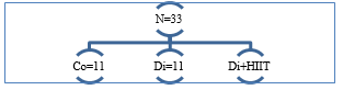 Figure 1. Distribution of research groups. Co: Control group; Di: Diabetic group; Di + HIIT: Diabetic + HIIT group |
In the second week, when the rats were well familiarized with the pool, they were trained to the interval format by being taken out of the water several times after one minute of swimming using a rest plate and then placed back in the water. Forty-eight hours after the last familiarization session, the rats in the training group first performed a 5-minute warm-up exercise and then the main HIIT swimming workout, which consisted of 14 repetitions of 20-second swimming exercises with 10 seconds of rest between each repetition. At the end of each training session, a 5-minute cool-down was applied, and then all rats were dried and returned to their cages. This exercise program was performed for eight weeks (Three days per week, on alternate days). In the load-interval training, the weight applied in the first week was 7% of the body weight of each rat and was increased by 1% each week so that in the final (Eighth) week the rats swam with a weight equivalent to 14% of their body weight tied to the root of their tails. Training was performed in the evening. During the intervention period, the control group did not perform any training program (13). For the load, a small weighted bead attached to the tail by a band (Tail band) was used. Before each session, the weight was adjusted to the body weight of each rat using a scale. Water depth was at least twice the body length of the rats, and its temperature was controlled at a constant 30 ± 0.5 °C throughout all sessions by a digital thermometer. Under supervision, the rats were allowed to swim at their natural speed; the researcher simply monitored them and did not force or stimulate increased speed. Animal fatigue was assessed by observing swimming behavior, stopping, diving, or attempting to escape; additional rest was given in case of severe fatigue. During the first one to two weeks, the rats adapted to the protocol and were able to complete all repetitions without stopping.
Research protocol
To induce diabetes, streptozotocin (STZ; 60 mg/kg; Sigma-Aldrich, USA; ≥98% purity by HPLC) dissolved in 0.1 M citrate buffer (pH 4.5) was injected subcutaneously between the two ears. Seven days after injection, blood glucose was measured, and rats with blood glucose levels higher than 250 mg/dL were selected as diabetic samples. Blood glucose was measured before STZ injection and on days 0, 7, 14, 21, and 35. Ketamine and xylazine were used to anesthetize the animals at doses of 80 mg/kg and 10 mg/kg, respectively, administered intraperitoneally (i.p.). This dose produced deep and stable anesthesia for surgical interventions and cardiac biopsy. After sampling, hearts were removed and stored at −80 °C until RNA extraction. To examine the expression of vascular VEGF-A and hypoxia-inducible factor genes, total cellular RNA was extracted using an RNA extraction column kit (FavorPrep™ Tissue Total RNA Mini Kit) according to the manufacturer's instructions.
Sampling was performed 48 hours after the last training session and after an overnight fast. First, the rats were anesthetized, the chest was opened, and blood was taken directly from the heart to minimize trauma. Heart muscle was separated under sterile conditions. Blood samples taken directly from the heart were collected and stored in Falcon tubes.
Real-time PCR protocol
RNA extraction
Total RNA was extracted from heart tissue samples (Approximately 50 mg per sample) stored at -80 °C using a commercial RNA isolation kit (RNX-Plus Synaclone). The tissue was homogenized in 1 mL TRIzol using a tissue homogenizer, followed by phase separation with chloroform (0.2 mL per 1 mL TRIzol). The aqueous phase was collected, and RNA was precipitated with isopropanol, washed with 75% ethanol, and resuspended in nuclease-free water.
RNA concentration and purity were assessed using a spectrophotometer (NanoDrop 2000, Thermo Fisher Scientific) at 260/280 nm, and an A260/A280 ratio of 1.8–2.0 was ensured. RNA integrity was confirmed by 1% agarose gel electrophoresis, which showed intact 18S and 28S ribosomal RNA bands.
cDNA synthesis
First-strand complementary DNA (cDNA) was synthesized from 1 μg of total RNA using a reverse transcription kit (iScript cDNA Synthesis Kit, Bio-Rad). The reaction mixture (20 μL) contained 1 μg of RNA, 4 μL of 5× iScript reaction mix, 1 μL of iScript reverse transcription enzyme, and nuclease-free water. Reverse transcription was performed in a thermal cycler (T100 Thermal Cycler, Bio-Rad) with the following conditions: 5 min at 25 °C (Priming), 20 min at 46 °C (Reverse transcription), and 1 min at 95 °C (Enzyme inactivation). The synthesized cDNA was diluted 1:10 with nuclease-free water and stored at -20 °C until analysis.
Primer design
Primers for vascular endothelial growth factor-A (VEGF-A), hypoxia-inducible factor-1α (HIF-1α), and a reference gene (e.g., GAPDH or β-actin) were designed using Primer-BLAST (NCBI) to ensure specificity and optimal amplification efficiency. Primers were synthesized by a commercial provider (e.g., Integrated DNA Technologies). Primer specificity was verified by melting curve analysis and agarose gel electrophoresis, confirming single amplicon production.
Real-time PCR
Quantitative PCR was performed using a real-time PCR system (CFX96 Touch, Bio-Rad). Each 20 μL reaction contained 10 μL of 2× SYBR Green Master Mix (PowerUp SYBR Green Master Mix, Thermo Fisher Scientific), 0.5 μM of each forward and reverse primer, 2 μL of diluted cDNA, and nuclease-free water. Cycling conditions included initial denaturation at 95 °C for 2 min, followed by 40 cycles of denaturation at 95 °C for 15 s, annealing at 60 °C for 30 s, and extension at 72 °C for 30 s. Melting curve analysis (65-95 °C, 0.5 °C increments) was performed after amplification to confirm amplicon specificity. All reactions were performed in triplicate, and no-template controls were included to detect contamination or nonspecific amplification.
Data analysis
Relative gene expression was calculated using the 2^ (-ΔΔCt) method. The cycle threshold (Ct) values of VEGF-A and HIF-1α were normalized to obtain ΔCt values relative to the reference gene (GAPDH). The ΔCt of the untrained diabetic group served as a calibrator for comparison of relative expression. Amplification efficiency was determined using standard curves generated from serial dilutions of cDNA, and efficiencies of 90–110% were ensured for all primer pairs. Data are expressed as mean ± standard deviation. The reference gene was GAPDH.
Extraction of HIF1-α RNA by column method
Left ventricular tissues were removed immediately after euthanasia, frozen in liquid nitrogen, and stored at -80 °C until processing. RNA extraction was performed using a column method. Frozen heart tissues were mechanically ground in liquid nitrogen in a mortar to obtain a fine powder for homogenization. The homogenized tissue was resuspended in 1 mL of RB lysis buffer (Containing β-mercaptoethanol) to lyse cells and stabilize RNA. The homogenate was centrifuged for 5 min at 4 °C at 12,000 rpm to remove debris. The supernatant was transferred to a filter column in a 2 mL collection tube and centrifuged for 2 min at 14,000 rpm to remove residual debris. The clear lysate was collected in a new microcentrifuge tube. The lysate was mixed with an equal volume of 70% ethanol and vortexed thoroughly to precipitate RNA. The mixture was transferred to an RNA-binding spin column and centrifuged for 1 min at 10,000 rpm. The flow-through was discarded, and the column was washed twice with RW1 buffer (700 μL) and RPE buffer (500 μL); after each wash, it was centrifuged for 1 min at 10,000 rpm to remove impurities. RNA was eluted by adding 30-50 μL of RNase-free water to the column, incubating for 1 min, and centrifuging at 10,000 rpm for 1 min. The washed RNA was stored at -80 °C. RNA quality was assessed by agarose gel electrophoresis. RNA samples (2 μg) were loaded on a 1.5% agarose gel stained with ethidium bromide. The presence of 28S and 18S ribosomal RNA bands (With an intensity ratio of ~2:1) confirmed RNA integrity.
cDNA synthesis
cDNA was synthesized from 1 μg of total RNA using the Fermentas cDNA synthesis kit (K1621) according to the manufacturer's instructions. The RNA was mixed with 1 μL of random hexamer primers and RNase-free water to a total volume of 12 μL. The mixture was incubated for 5 min at 65 °C to denature RNA secondary structures. A master mix containing 4 μL of 5× reaction buffer, 1 μL of RiboLock RNase inhibitor, 2 μL of 10 mM dNTP mix, and 1 μL of RevertAid Reverse Transcriptase was added. Reverse transcription was performed in a thermal cycler (25 °C for 10 min, 42 °C for 60 min, and 70 °C for 5 min to inactivate the enzyme). The resulting cDNA was stored at -20 °C.
Real-time PCR for gene expression analysis
VEGF-A gene expression was measured using quantitative real-time PCR (qRT-PCR). β2-microglobulin (β2m) and glyceraldehyde-3-phosphate dehydrogenase (GAPDH) genes were used as internal controls for normalization, and primer design was performed using ID v7.8 software. Primer sequences were validated using BLAST, and PCR efficiency was calculated using a standard curve generated from serial dilutions of cDNA (10-1000 ng). Primer efficiency ranged from 90-110%. Reactions were performed in a 20 μL volume containing 25 ng cDNA, 10 μL RealQ 2× Master Mix Green Dye (Ampliqon, Germany), 0.5 μM of each forward and reverse primer, and nuclease-free water. Thermal cycling conditions were: initial denaturation at 95 °C for 10 min, followed by 40 cycles of 95 °C for 15 s and 60 °C for 60 s. Melting curve analysis (65-95 °C) was performed to confirm amplicon specificity. Data analysis and relative gene expression were calculated using the 2^(-ΔΔCt) method. VEGF-A Ct values were normalized to the mean Ct values of β2m and GAPDH.
VEGF-A) Vascular Endothelial Growth Factor) measurement by ELISA
VEGF-A protein levels in heart tissue homogenates were determined using a sandwich enzyme-linked immunosorbent assay (ELISA) with a sensitivity of <2 pg/mL, according to the manufacturer's instructions (MyBioSource, Catalog number: MBS2881654 Rat Hypoxia-Inducible Factor 1 Alpha). For sample preparation, frozen heart tissues (100 mg) were homogenized in 1 mL of cold phosphate-buffered saline (PBS, pH 7.4) containing protease inhibitors (1 mM PMSF, 1 μg/mL aprotinin) using a tissue homogenizer. The homogenates were centrifuged at 12,000 rpm for 10 min at 4 °C, and the supernatant was collected for analysis. Total protein concentrations were determined using the Bradford method to normalize VEGF-A levels. A 96-well microplate was precoated with a monoclonal antibody specific for rat VEGF-A and incubated overnight at 4 °C. The plate was washed three times with buffer (0.05% Tween-20 in PBS) to remove unbound antibody. The wells were blocked with 1% bovine serum albumin (BSA) in PBS for 1 h at room temperature to prevent nonspecific binding. Tissue homogenates (100 μL) and serial dilutions of recombinant rat VEGF-A (For standard curve, 0-1000 pg/mL) were added to the wells in duplicate and incubated for 2 h at room temperature. After four washes, a biotinylated detection antibody specific for VEGF-A was added and incubated for 1 h at room temperature. The plate was washed again, and a streptavidin-horseradish peroxidase conjugate was added for 30 min. After a final wash, tetramethylbenzidine substrate was added, and the reaction was incubated for 20 min in the dark. The reaction was stopped with 2 N sulfuric acid, and absorbance was measured at 450 nm (Corrected to 570 nm) using a microplate reader (BioTek). VEGF-A concentrations (Picograms per milliliter) were calculated from the standard curve and normalized to total protein (Picograms per milligram of protein). Positive controls (Known concentrations of VEGF-A) and negative controls (PBS only) were included to validate assay performance. The standard curve was fitted using a four-parameter logistic regression model to ensure linearity and precision. All samples were tested in duplicate, and intra-assay and inter-assay coefficients of variation were <5% and <10%, respectively.
To minimize bias, heart tissue samples were analyzed in a blinded manner. The rats were divided into groups by simple randomization, and extraction and measurement protocols (ELISA) were performed uniformly for all samples.
Statistical analysis
Gene expression results are presented as mean ± standard deviation (SEM) of three independent replicates. The mean mRNA expression level of treated samples was compared with the mean of control samples (=1). The levels of the studied variables, including VEGF-A and HIF-1α, are reported as mean ± standard deviation. All data were first checked for normality using the Shapiro–Wilk test. Levene’s test was used to evaluate the equality of variances, with a P-value greater than 0.05 indicating homogeneity. and comparisons between the groups were performed using one-way analysis of variance (One-way ANOVA). If significant differences were observed, Tukey's post hoc test was used for pairwise comparisons. All analyses were performed using SPSS version 19 software; Statistical significance level was 0.05.
Results
The mean ± SD of VEGF-A and HIF-1α gene expression were measured in the three groups. VEGF-A expression was increased in the diabetic group subjected to HIIT, reaching levels similar to those of the control group. Conversely, HIF-1α expression was reduced in the diabetic + HIIT group, approaching control group levels (Figures 2 and 3).
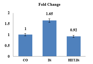 Figure 2. Comparison of descriptive characteristics of VEGF-A gene expression (Mean ± SD) The results of the mean and standard deviation showed that HIF-1α decreased in the HIIT group.  Figure 3. Comparison of descriptive characteristics of HIF-1α gene expression (Mean ± SD) The results of the mean and standard deviation showed that HIF-1α increased in the HIIT group. |
Tukey's post-hoc test revealed a significant increase in VEGF-A levels in the diabetic + HIIT group compared to the diabetic group (P-Value = 0.000). Additionally, a significant difference was noted between the diabetic group and the control group (P-Value = < 0.001). Regarding HIF-1α, the mean and standard deviation indicated an elevation in the diabetic group. Tukey's post-hoc test confirmed a significant increase in HIF-1α in the diabetic group compared to the diabetic + HIIT group (P-Value = 0.026), as well as a significant difference between the diabetic group and the control group (P-Value = 0.001) (Table 1).
|
Table 1. Results of one-way analysis of variance and Tukey post hoc test of dependent variables of the research
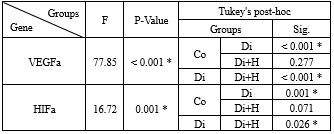 *: P-Value < 0.05 |
The large effect size for VEGF-A (η² = 0.945) highlights a significant impact of HIIT training on this gene's expression. Similarly, a large effect size for HIF-1α (η² = 0.758) indicates a strong influence of the interventions on this gene.
These results suggest that HIIT can modify the expression of key genes involved in angiogenesis (VEGF-A) and hypoxic response (HIF-1α). While these molecular changes are recognized as potential mechanisms, they do not guarantee improvements in cardiac function, such as increased cardiac output, enhanced ejection fraction, or reduced fibrosis. Elevated VEGF-A and HIF-1α are typically linked to angiogenesis stimulation, improved oxygenation, and regulation of cardiac metabolism in diabetic conditions, which could over time enhance blood supply, reduce tissue ischemia, and improve the heart's ability to handle stress. Thus, these findings provide a foundation for further research at the functional level, including echocardiography, functional tests, or histological analyses. Moreover, the observed gene expression changes serve as primary molecular indicators, pointing to the beneficial effects of HIIT on angiogenic mechanisms and hypoxic responses in the diabetic heart. However, to validate functional and clinical outcomes, additional studies directly evaluating cardiac function and myocardial structure are essential.
Discussion
This study demonstrated that HIIT significantly increased HIF-1α gene expression in diabetic animals compared with diabetic controls. These findings align with previous evidence indicating that exercise training can upregulate HIF-1α expression. Soltani et al. (2019) reported increased HIF-1α expression following aerobic training in female mice (17), and De Carvalho et al. (2022) observed a similar effect in cardiac tissue of Wistar rats under aerobic training and hypoxic conditions (18).
Inconsistencies across studies may be explained by differences in exercise type, intensity, duration, and animal models. Furthermore, the long-term activity of HIF-1α after chronic training remains unclear, likely due to its transient regulation, as mRNA expression and target gene activity are often used as indirect markers of HIF-1α function (19).
Previous studies have also shown that endurance training enhances HIF-1α expression in skeletal muscle and lung tissue (19). Zeng et al. (2025) suggested that HIF-1α transcription represents an early adaptive response to hypoxia, regulating glycolysis and angiogenesis (20). Similarly, Song et al. demonstrated that elevated HIF-1α during exercise can act as an upstream regulator of miR-126, thereby promoting angiogenesis via the eNOS/AKT/PI3K and MAPK pathways and improving cardiac function (21).
In this study, diabetes was induced using STZ in gerbils. However, variable responses to STZ should be acknowledged, as some animals exhibit greater cellular damage than others, which may affect the observed outcomes.
Another important finding of the present study was a significant increase in serum VEGF levels in the HIIT group. Exercise-induced ischemia appears to activate angiogenic pathways by re-stimulating cardiac VEGF, which promotes coronary capillary growth and improves myocardial perfusion. These adaptations contribute to enhanced left ventricular function, improved myocyte contractility in response to adrenergic stimulation, and prevention of adverse cardiac remodeling. The observed improvements in systemic hemodynamics and physical performance further indicate a positive overall effect of exercise training (22).
At the molecular level, exercise activates signaling responses through free radicals such as nitric oxide. VEGF plays a central role in hypoxia-induced cell proliferation by promoting adenosine production and receptor activation in endothelial cells. In addition, hypoxic conditions upregulate VEGF and its receptor VEGFR-1 via HIF-1α binding to the hypoxia response element (23). Supporting these findings, Kheradmand et al. (2023) reported a significant increase in VEGF gene expression following endurance and interval training in rats (24), while Monjezi et al. (2024) demonstrated an increase in VEGFR-1 concentration in muscle tissue after physical activity (25). Similarly, Soori et al. showed that treadmill training in type 1 diabetic rats increased VEGF expression in cardiac muscle, whereas VEGF levels were markedly reduced in inactive diabetic animals (26). These results suggest that the elevated VEGF levels observed in our study may reflect a protective role of HIIT against cardiac fibrosis and its complications through enhanced angiogenesis and vascular remodeling (27).
However, not all studies have confirmed these effects. Song et al. (2024) found no significant changes in serum VEGF levels in overweight men aged 50-60 years after a long-term training program (Running and cycling), possibly due to adaptive responses or differences in sampling (Plasma vs. serum) (28).
Likewise, Salgueiro et al. (2017) reported that endurance training reduced VEGF, and no significant differences were observed between exercise and supplementation groups in myocardial ischemia, potentially due to the concurrent use of L-arginine supplements (29).
In addition to its central role in angiogenesis, VEGF-A contributes to improved tissue oxygenation by promoting vascular endothelial cell proliferation, enhancing vascular permeability, and increasing blood supply to target organs. HIF-1α, which is activated under hypoxic conditions, regulates the expression of hypoxia-responsive genes, including VEGF-A. Through its role in controlling energy metabolism, angiogenesis, and cell survival, HIF-1α facilitates cardiac adaptation to metabolic stress induced by diabetes (30).
HIIT consists of short bouts of intense exercise interspersed with periods of rest or low-intensity activity. This pattern imposes controlled metabolic and hypoxic stress on tissues, which can activate signaling pathways that upregulate the expression of both VEGF-A and HIF-1α, thereby promoting angiogenesis, improving tissue oxygenation, and enhancing cardiac adaptation (31).
During HIIT, the high intensity of exercise increases cardiac oxygen demand, which may lead to localized hypoxia. Under these conditions, HIF-1α is stabilized because hypoxia inhibits its degradation by prolyl hydroxylases. The activated HIF-1α then translocates to the nucleus and induces the expression of target genes, including VEGF-A, thereby promoting angiogenesis and enhancing tissue oxygenation (32). In the diabetic heart, hypoxic responses may be impaired due to oxidative stress and disrupted signaling pathways. HIIT can reactivate these pathways by inducing moderate levels of reactive oxygen species (ROS), which act as molecular signals. ROS can activate signaling cascades such as MAPK (Mitogen-activated protein kinases) and PI3K/Akt, both of which play critical roles in the regulation of HIF-1α and VEGF-A, thereby promoting angiogenesis and improving cardiac adaptation (33).
In diabetes, chronic oxidative stress impairs the heart’s adaptive responses. HIIT can modulate ROS production to stimulate antioxidant defenses, including increased activity of enzymes such as superoxide dismutase and catalase. Simultaneously, HIIT enhances the expression of protective genes, including VEGF-A, thereby improving cardiac resilience and promoting angiogenesis (34). HIIT also activates AMPK (AMP-activated protein kinase), a critical metabolic sensor. AMPK can directly stimulate VEGF-A expression or indirectly enhance it through interaction with HIF-1α. In addition, AMPK improves mitochondrial metabolism, which is often impaired in the diabetic heart. Since diabetes reduces AMPK activity by disrupting glucose and lipid metabolism, HIIT can reactivate this pathway, thereby improving myocardial perfusion and overall cardiac function (35).
Diabetes is associated with chronic inflammation, which can suppress the expression of VEGF-A and HIF-1α. HIIT creates a favorable molecular environment by reducing pro-inflammatory markers such as TNF-α and IL-6 while increasing anti-inflammatory cytokines like IL-10. This reduction in inflammation enhances endothelial function and amplifies the tissue response to angiogenic signals, thereby supporting vascular remodeling and cardiac adaptation (35).
Finally, HIIT may regulate the expression of VEGF-A and HIF-1α through epigenetic mechanisms, such as reduced DNA methylation or histone modifications, which increase the accessibility of transcription factors to gene promoters. In diabetes, negative epigenetic changes suppress the expression of protective genes, contributing to impaired cardiac function. HIIT can potentially reverse these effects. In the diabetic heart, multiple factors-including oxidative stress, chronic inflammation, impaired energy metabolism, and reduced angiogenesis-contribute to decreased cardiac performance. HIIT counteracts these maladaptive processes by enhancing angiogenesis and metabolic capacity. Activation of HIF-1α and AMPK improves glucose and lipid metabolism, while increased expression of VEGF-A and HIF-1α protects the heart against ischemia–reperfusion injury and supports overall cardiac adaptation.
The intensity and duration of HIIT, the type of diabetes (Type 1 or 2), and the use of supplementation (If any) can influence the results. Also, the long-term effects of HIIT on the diabetic heart need further investigation.
For future directions, it is suggested to investigate the direct effect of VEGF-A and HIF-1α on cardiac function and the role of supplementation (Such as antioxidants or metformin) in enhancing these pathways.
Conclusion
HIIT likely increases VEGF-A and HIF-1α expression in the diabetic heart through induction of local hypoxia, activation of AMPK and ROS pathways, reduction of inflammation, and epigenetic changes. These changes may contribute to improved angiogenesis, energy metabolism, and protection against ischemia, which are critical for the diabetic heart. The available scientific literature supports these mechanisms, but physiological data and additional studies are needed to confirm a direct link to cardiac function.
Acknowledgement
We thank all experts who collaborated in the animal work, dissection, histology, and experiments.
Funding sources
This work was funded by the authors' expenses.
Ethical statement
All animal experiments were conducted in accordance with the National Institutes of Health (NIH) Guidelines for the Care and Use of Laboratory Animals. The study protocol was reviewed and approved by the Ethics Committee of the Faculty of Medicine, Varamin Pishva Branch, Islamic Azad University, under the ethical approval code IR.IAU.VARAMIN.REC.1402.048. Every effort was made to minimize animal suffering and to reduce the number of animals involved.
Conflicts of interest
The authors declare no conflicts of interest.
Author contributions
Conceptualization and Methodology: F.N. and E.J.; Formal analysis and Research: F.N. and E.J.; Writing-Original draft preparation: F.N.; Writing-Review and Editing: F.N. All authors have read and approved the published version of the manuscript.
Data availability statement
The datasets generated and analyzed during the current study can be obtained from the corresponding author upon a reasonable request.
Editorial: Original article |
Subject:
Health
Received: 2025/03/16 | Accepted: 2025/09/28 | Published: 2025/09/29
Received: 2025/03/16 | Accepted: 2025/09/28 | Published: 2025/09/29
References
1. Zheng S, Ni J, Li Y, Lu M, Yao Y, Guo H, et al. 2-Methoxyestradiol synergizes with Erlotinib to suppress hepatocellular carcinoma by disrupting the PLAGL2-EGFR-HIF-1/2α signaling loop. Pharmacol Res. 2021;169:105685. [View at Publisher] [DOI] [PMID] [Google Scholar]
2. Batrakoulis A, Jamurtas AZ, Fatouros IG. High-Intensity Interval Training in Metabolic Diseases: Physiological Adaptations. ACSMs Health Fit J. 2021;25(5):54-9. [View at Publisher] [DOI] [Google Scholar]
3. Syeda UA, Battillo D, Visaria A, Malin SK. The importance of exercise for glycemic control in type 2 diabetes. Am J Med Open. 2023;9:100031. [View at Publisher] [DOI] [PMID] [Google Scholar]
4. Bayat M, Alaee M, Akbari A, Sadegh M, Latifi SA, Parastesh M, et al. A comparative study of the antidiabetic effect of two training protocols in streptozotocin-nicotinamide diabetic rats. Horm Mol Biol Clin Investig. 2020;41(2). [View at Publisher] [DOI] [PMID] [Google Scholar]
5. Yang Ch, Zhong ZF, Wang Sh-P, Vong Ch-T, Yu B, Wang Y-T. HIF-1: structure, biology and natural modulators. Chin J Nat Med. 2021;19(7):521-7. [View at Publisher] [DOI] [PMID] [Google Scholar]
6. Drozdovska S, Zanou N, Lavier J, Mazzolai L, Millet GP, Pellegrin M. Moderate Effects of Hypoxic Training at Low and Supramaximal Intensities on Skeletal Muscle Metabolic Gene Expression in Mice. Metabolites. 2023;13(10):1103. [View at Publisher] [DOI] [PMID] [Google Scholar]
7. Villamil-Parra W, Cristancho-Mejía É, Ramon Torrella J, Mancera-Soto EM. Effects of a physical exercise program on HIF-1α in people with Chronic Obstructive Pulmonary Disease living at high altitude: study protocol for a clinical trial. Trials. 2023;24(1):698. [View at Publisher] [DOI] [PMID] [Google Scholar]
8. Emami Nejad A, Najafgholian S, Rostami A, Sistani A, Shojaeifar S, Esparvarinha M,et al. The role of hypoxia in the tumor microenvironment and development of cancer stem cell: a novel approach to developing treatment. Cancer Cell Int. 2021;21(1):62. [View at Publisher] [DOI] [PMID] [Google Scholar]
9. DA Silva BVC, Branco DBT, Ide BN, Marocolo M, DE Souza HLR, Arriel RA, et al. Comparison of High-Volume and High-Intensity Upper Body Resistance Training on Acute Neuromuscular Performance and Ratings of Perceived Exertion. Int J Exerc Sci. 2020;13(1):723-3. [View at Publisher] [DOI] [PMID] [Google Scholar]
10. Zokaei A, Ghahramani M, Bolouri G, Nassiri Avanaki M. Comparison of Continuous and Periodic Exercise on Serum Nitric Oxide Level and Vascular Endothelial Growth Factor in Old Rats. Mljgoums. 2023;17(2):20-25. [View at Publisher] [DOI] [Google Scholar]
11. Ghahramani M, Razavi Majd Z. The Effect of Physical Activity on VEGF and HIF-1 Signaling. J Clin Res Paramed Sci. 2020;9(2):e98493. [View at Publisher] [DOI] [Google Scholar]
12. Luo Z, Tian M, Yang G, Tan Q, Chen Y, Li G, et al. Hypoxia signaling in human health and diseases: implications and prospects for therapeutics. Signal Transduct Target Ther. 2022;7(1):218. [View at Publisher] [DOI] [PMID] [Google Scholar]
13. Amirazodi M, Mehrabi A, Rajizadeh MA, Bejeshk MA, Esmaeilpour K, Daryanoosh F, et al. The effects of combined resveratrol and high intensity interval training on the hippocampus in aged male rats: An investigation into some signaling pathways related to mitochondria. Iran J Basic Med Sci. 2022;25(2):254-62. [View at Publisher] [DOI] [PMID] [Google Scholar]
14. Su Z, Chen L, Lu M, Li J, Qiu L, Shi L. Peripheral blood HIF-1α levels: a study on their predictive ability for early-stage diabetic retinopathy. Endocr Connect. 2025;14(5):e250004. [View at Publisher] [DOI] [PMID] [Google Scholar]
15. Cerychova R, Pavlinkova G. HIF-1, Metabolism, and Diabetes in the Embryonic and Adult Heart. Front Endocrinol (Lausanne). 2018;9:460. [View at Publisher] [DOI] [PMID] [Google Scholar]
16. Thangarajah H, Vial IN, Grogan RH, Yao D, Shi Y, Januszyk M, et al. HIF-1alpha dysfunction in diabetes. Cell Cycle. 2010;9(1):75-9. [View at Publisher] [DOI] [PMID] [Google Scholar]
17. Soltani R, Kordi MR, Gaeini AA, Nuri R. The effects of 8 weeks aerobic training on HIF-1α, miR-21 and VEGF gene expression in female Balb/c with breast cancer. Yafte. 2019;21(1):63-74. [View at Publisher] [Google Scholar]
18. De Carvalho CD, Valentim RR, Navegantes LC, Papoti M. Comparison between low, moderate, and high intensity aerobic training with equalized loads on biomarkers and performance in rats. Sci Rep. 2022;12(1):18047 [View at Publisher] [DOI] [PMID] [Google Scholar]
19. Furrer R, Handschin C. Molecular aspects of the exercise response and training adaptation in skeletal muscle. Free Radic Biol Med. 2024;223:53-68. [View at Publisher] [DOI] [PMID] [Google Scholar]
20. Zeng Y, Tao Y, Du G, Huang T, Chen S, Fan L, et al. Advances in the mechanisms of HIF-1α-enhanced tumor glycolysis and its relation to dedifferentiation. Prog Biophys Mol Biol. 2025:197:1-10 [View at Publisher] [DOI] [PMID] [Google Scholar]
21. Song W, Liang Q, Cai M, Tian Z. HIF-1α-induced up-regulation of microRNA-126 contributes to the effectiveness of exercise training on myocardial angiogenesis in myocardial infarction rats. J Cell Mol Med. 2020;24(22):12970-9. [View at Publisher] [DOI] [PMID] [Google Scholar]
22. Kirsch M, Iliou M-C, Vitiello D. Hemodynamic Response to Exercise Training in Heart Failure With Reduced Ejection Fraction Patients. Cardiol Res. 2024;15(1):18-28. [View at Publisher] [DOI] [PMID] [Google Scholar]
23. Lucchesi M, Di Marsico L, Guidotti L, Lulli M, Filippi L, Marracci S, Dal Monte M. Hypoxia-Dependent Upregulation of VEGF Relies on β3-Adrenoceptor Signaling in Human Retinal Endothelial and Müller Cells. Int J Mol Sci. 2025;26(9):4043. [View at Publisher] [DOI] [PMID] [Google Scholar]
24. Kheradmand S, Asad MR, Mir Javadi R, Kheradmand N, Fashi M. Influence of Exercise Intensity on the Expression of Angiogenesis-Related Genes in the Hearts of Male Rats. J Basic Res Med Sci. 2023;10(4):43-53 [View at Publisher] [Google Scholar]
25. Monjezi M, Barari A, Abdi A. The Impact of Specific Physical Training on FGF-2 and VEGF-A Expression in Patients Post-Coronary Artery Bypass. J Bas Res Med Sci. 2024;11(3):56-65. [View at Publisher] [Google Scholar]
26. Soori R, SHarafi dehrahm F, CHoobine S, Valipour dehnou V. Effect of endurance training on VEGF protein level in tissue of cardiac muscle in STZ-induced diabetic Wistar rats. Yafte. 2018;20(3):110-24. [View at Publisher] [Google Scholar]
27. Yazdani F, Shahidi F, Karimi P. The effect of 8 weeks of high-intensity interval training and moderate-intensity continuous training on cardiac angiogenesis factor in diabetic male rats. J Physiol Biochem. 2020;76(2):291-9. [View at Publisher] [DOI] [PMID] [Google Scholar]
28. Song BX, Azhar L, Koo GKY, Marzolini S, Gallagher D, Swardfager W, et al. The effect of exercise on blood concentrations of angiogenesis markers in older adults: a systematic review and meta-analysis. Neurobiol Aging. 2024:135:15-25. [View at Publisher] [DOI] [PMID] [Google Scholar]
29. Salgueiro RB, Gerlinger-Romero F, Guimarães-Ferreira L, de Castro Barbosa T, Nunes MT. Exercise training reverses the negative effects of chronic L-arginine supplementation on insulin sensitivity. Life Sci. 2017;191:17-23. [View at Publisher] [DOI] [PMID] [Google Scholar]
30. Zhang J, Yao M, Xia S, Zeng F, Liu Q. Systematic and comprehensive insights into HIF-1 stabilization under normoxic conditions: implications for cellular adaptation and therapeutic strategies in cancer. Cell Mol Biol Lett. 2025;30(1):2. [View at Publisher] [DOI] [PMID] [Google Scholar]
31. Hall B, Żebrowska A, Sikora M, Siatkowski S, Robins A. The Effect of High-Intensity Interval Exercise on Short-Term Glycaemic Control, Serum Level of Key Mediator in Hypoxia and Pro-Inflammatory Cytokines in Patients with Type 1 Diabetes-An Exploratory Case Study. Nutrients. 2023;15(17):3749. [View at Publisher] [DOI] [PMID] [Google Scholar]
32. Figarella K, Kim J, Ruan W, Mills T, Eltzschig HK, Yuan X. Hypoxia-adenosine axis as therapeutic targets for acute respiratory distress syndrome. Front Immunol. 2024;15:1328565. [View at Publisher] [DOI] [PMID] [Google Scholar]
33. Chen H, Guo L. Exercise in Diabetic Cardiomyopathy: Its Protective Effects and Molecular Mechanism. Int J Mol Sci. 2025;26(4):1465. [View at Publisher] [DOI] [PMID] [Google Scholar]
34. Firouzyar F, Shamlou Kazemi S, Hemmati Afif A. The Effect of High-Intensity Interval Training on Antioxidant Factors in Women with Type 2 Diabetes. JEHS. 2022;2(3):1-14. [View at publisher] [DOI] [Google Scholar]
35. Zhao L, Hu H, Zhang L, Liu Z, Huang Y, Liu Q, et al. Inflammation in diabetes complications: molecular mechanisms and therapeutic interventions. MedComm (2020). 2024;5(4):e516. [View at publisher] [DOI] [PMID] [Google Scholar]
Send email to the article author
| Rights and permissions | |
 |
This work is licensed under a Creative Commons Attribution-NonCommercial 4.0 International License. |