Volume 13, Issue 4 (12-2025)
Jorjani Biomed J 2025, 13(4): 27-33 |
Back to browse issues page



Download citation:
BibTeX | RIS | EndNote | Medlars | ProCite | Reference Manager | RefWorks
Send citation to:


BibTeX | RIS | EndNote | Medlars | ProCite | Reference Manager | RefWorks
Send citation to:
Jalili Sadrabad M, Sohanian S, Owlia F, Karimyan E. Association between periodontal indices and rheumatoid arthritis based on the status of disease control: A cross-sectional pilot study. Jorjani Biomed J 2025; 13 (4) :27-33
URL: http://goums.ac.ir/jorjanijournal/article-1-1103-en.html
URL: http://goums.ac.ir/jorjanijournal/article-1-1103-en.html


1- Department of Oral and Maxillofacial Medicine, School of Dentistry, Semnan University of Medical Sciences, Semnan, Iran
2- Department of Oral and Maxillofacial Pathology, School of Dentistry, Semnan University of Medical Sciences, Semnan, Iran
3- Department of Oral and Maxillofacial Medicine, School of Dentistry, Shahid Sadoughi University of Medical Science, Yazd, Iran ,f.owlia@ssu.ac.ir
4- Department of Restorative Dentistry, School of Dentistry, Shahid Sadoughi University of Medical Science, Yazd, Iran
2- Department of Oral and Maxillofacial Pathology, School of Dentistry, Semnan University of Medical Sciences, Semnan, Iran
3- Department of Oral and Maxillofacial Medicine, School of Dentistry, Shahid Sadoughi University of Medical Science, Yazd, Iran ,
4- Department of Restorative Dentistry, School of Dentistry, Shahid Sadoughi University of Medical Science, Yazd, Iran
Full-Text [PDF 534 kb]
(379 Downloads)
| Abstract (HTML) (3220 Views)
Results
Totally, 50 RA patients consisted of 5 males and 45 females with an age range of 23 to 65. Table 1 presents the frequency distribution of demographic data, laboratory tests, and clinical parameters across the study groups. The majority of patients were female (n=45, 90%), while 5 (10%) were male. The mean age of female patients was 44.4 ± 10.8 years (Range: 23-65), while that of male patients was 43.4 ± 4.5 years (Range: 39-48). There was no statistically significant difference between groups in terms of gender.
There was no significant difference in demographic characteristics or periodontal parameters among the groups. ESR was the only laboratory parameter that differed significantly among the groups (P-Value=0.016).
The mean hemoglobin level in the Inactive RA group was 12.86 ± 1.85 g/dL (Range: 9.1-16.3 g/dL). In the active RA group, the mean hemoglobin level was 12.95 ± 1.41 g/dL (Range: 10-15 g/dL). The mean hemoglobin level in the recurrent active RA group was 13.16±1.23 g/dl, with a range of 9.9 to 15.3 g/dl. Statistical analysis showed no significant difference in hemoglobin levels among the groups (P-Value=0.724) (Table 1). Based on CAL, 12(24%) patients had mild periodontitis, 25(50%) had moderate periodontitis, and 13(26%) had severe periodontitis.
Furthermore, differences in periodontal parameters between age subgroups were evaluated across all three groups. The Wilcoxon test was used to assess the differences between ESR, CAL, PD, and TL in the age subgroups (Table 2). None of the periodontal indices, including ESR, differed significantly between the age subgroups (≤40 vs. >40 years) across any of the three study groups. It indicates that although the clinical status of RA differed among groups, the periodontal indices did not differ significantly.
Discussion
Various studies have emphasized the existence of common genetic factors between RA and periodontitis (10-13,28). In humans, the regulatory genes of the monocytic cytokine response are located in the HLA-DR region of chromosome 5, within the TNF-β area. These genes are implicated in both chronic periodontitis and RA (10).
Previous studies have shown that peptidyl arginine deiminase (PAD) is involved in the pathogenesis of autoimmune damage in RA (29). It is produced by some human cells, such as lung cells, as well as by the microorganism Porphyromonas gingivalis, which supports the association between periodontitis and RA (30-32). The concentration of anti-citrullinated protein antibody (ACPA) and RF is elevated in the serum of patients with RA who are affected by chronic periodontitis (13). Porphyromonas gingivalis generates the ACPA in RA patients, suggesting a direct intersection between periodontitis and RA (33).
Some studies have considered CRP and ESR as components of DAS28 (Disease Activity Score) in their studies to classify RA patients (34,35). Despite this, a large United States registry of RA patients showed that acute phase reactant (APR) levels often do not correlate with disease activity as measured by joint counts and global assessments (36). Furthermore, ESR and CRP are not pathognomonic for RA activity, as they can be evaluated in all inflammatory conditions and infectious conditions (37). Therefore, clinical criteria formed the basis of classification. In 2020, both the European Federation of Periodontology and the American Academy of Rheumatology concluded that there was minimal evidence of a direct association between the two diseases (38).
Periodontitis is more prevalent in individuals in the earliest stages of seropositive RA (39). Kordtabar et al. reported that the gingival indices in RA patients who received anti-rheumatic drugs were significantly lower than in those who did not take medicine. The gingival index in patients with a history of consuming anti-rheumatic drugs was significantly lower than in those who were not drug consumers, showing the effect of the medications on the signs of inflammation (9).
The results of the current study showed no statistically significant difference in BOP, CAL, PD, and TL among different groups of RA patients. According to the analysis, ESR was the only parameter that differed significantly among the groups. The study’s recruitment setting may explain this observation. Since participants were selected exclusively from a rheumatology clinic, their chief complaint was inherently rheumatic disease rather than oral conditions. Patients experiencing oral manifestations secondary to rheumatic disease likely sought treatment at dental centers instead, which may have influenced the prevalence rates observed in this cohort. Smoking and diabetes, as important confounding variables, were excluded from this study to allow a more rigorous investigation of the possible relationship between these two chronic diseases.
The demographic characteristics of our participants were consistent with those of previous study, indicating that RA was more common in females (38). Methodological differences, particularly regarding age stratification, may account for the variations observed between our findings and previous studies. In the present study, the mean age of participants was 44 years. This aligns closely with the typical age of onset of approximately 40 years, when many autoimmune conditions, such as rheumatoid arthritis (RA), typically become clinically apparent. Consequently, 40 years was selected as the cut-off point for stratifying age groups (40). Furthermore, our sample size (N=50) differed from prior studies, which may have influenced statistical power. This study affirmed the findings of a previous study that showed no correlation between RA and periodontitis status (38). This discrepancy may be attributable to differences in ethnic groups and adjustments for confounding variables across different populations (41).
This study found no significant differences in periodontitis indices among RA groups, which is consistent with previous studies (38,39). In contrast, Khantisopon et al. (22) reported a strong relationship among Thai patients. This discrepancy may be attributed to the high prevalence of periodontitis in the general Thai population (22) or to the different classifications of RA patients in this study.
Almashni et al. (2025) concluded that while marginal bone loss and implant survival rates were comparable between RA and non-RA patients, the RA group exhibited a significantly higher plaque index (PI) (42). While our findings showed no significant difference in tooth loss among the study groups, a key distinction lies in the study population. All participants in the current study were RA patients, whereas Almashni et al. examined differences between RA patients and healthy individuals (42). Given the chronic nature of RA, all participants had been exposed to systemic inflammation for an extended duration, which may explain the lack of significant differences in this study between subgroups compared to studies with healthy controls (43). According to the scoping review, there was a close association between bone loss and inflamed joints as well as with CRP and ESR levels. They found a direct relationship between periodontitis and RA (44). The discrepancy between the two studies may be due to different methods of periodontal assessment and the limitation of the sample size, and insufficient statistical power.
Pischon et al. evaluated the association among RA, oral hygiene, and periodontitis in 57 patients with active RA and 57 healthy individuals. The results showed RA patients had more severe periodontitis than the control group (17). Finally, they reported RA patients had significantly higher CAL. They concluded that oral hygiene may only partially account for this relationship. In this study, oral health status was a confounding factor that was not assessed. Dissick et al. investigated the relationship between periodontitis and RA in 69 patients with RA and 35 healthy individuals, assessing their PD and panoramic radiographs (10). They reported that the prevalence of moderate to severe periodontitis in patients with RA was higher than in controls, independent of age, sex, smoking, or diabetes mellitus (10) Our study was consistent with theirs.
Although Kim et al. found no association between chronic periodontitis and rheumatoid arthritis in the Korean population, they concluded that RA was associated with tooth loss (TL) in younger adults (32). While many studies have discussed the relationship between these two diseases and found a significant relationship between CAL, PI, BOP, and PD (45,46). A study found no noteworthy relationship between periodontitis and RA, which is consistent with our findings (1). Wei et al. evaluated a novel target, cathepsin K (Ctsk)-mediated TLR9-related autophagy, during the progression of periodontitis in RA patients (47). Their study utilized a mouse model of periodontitis with RA. Small interfering RNA (siRNA) and CpG oligodeoxynucleotides (CpG ODN) were administered to macrophages. The results showed that RA can promote periodontitis bone destruction in the lesion area. Macrophage stimulation experiments confirmed the in vivo results. This study, the findings of which differ from ours, identified a novel role for Ctsk in TLR9-mediated autophagy to explain the interaction between periodontitis and RA (47).
Rodríguez-Lozano et al. evaluated the association between periodontitis severity and clinical activity in rheumatoid arthritis patients in a case-control study (48). Periodontal exposure was assessed using the following periodontal parameters: PI, BOP, PD, and CAL. They showed a significant association between periodontitis and RA with all periodontal parameters being significantly worse in RA patients than in controls. Periodontitis severity was significantly associated with RA disease activity (48). The inconsistency between their results and ours could be explained by the limited sample size, the wide age range of patients, and the use of different statistical tests.
Future research should assess the diagnosis of periodontitis, the presence of serum ACPA, and their association with the clinical manifestations of RA. The relationship between periodontitis treatment and the reduction of the risk of RA with ACPA positivity should also be examined (13).
Since the data interval was highly dispersed with respect to gender and age range, and the probability of detecting correlations with these variables was low, no significant relationship between different groups was found in the present study. Therefore, future studies with groups matched for periodontal status and with a narrower age range would yield definitive results.
In this study, with a clinical approach, hemoglobin was categorized using established clinical thresholds (WHO criteria for anemia: <12, 12-13, >13 g/dL) to ensure findings are directly interpretable for clinical decision-making. While the authors acknowledge the statistical implications, this approach prioritized clinical utility over continuous analysis in this pilot study.
Although TL in the recurrent active group (23.36%) was higher than in the inactive group (12.83%), there was no statistically significant difference. Due to RA-related disability, such as joint involvement, these patients may have difficulty maintaining good oral hygiene. Dentists play a vital role in achieving and maintaining optimal oral health through timely oral and dental examinations. Despite the many studies conducted on this topic, there is little evidence concerning a possible correlation between RA and periodontitis (39).
Despite its limitations, the study is important and novel in its new approach to stratifying RA patients for the design of future studies. The results showed no statistically significant difference in periodontal indices among the RA groups. Although routine oral health care may seem sufficient for these patients, a specialized protocol for RA patients is needed. This work can be a valuable foundation for the treatment of RA patients.
However, future studies should investigate ACPA and its association with periodontitis. The effect of periodontal treatment on RA parameters and RA duration since diagnosis is another relevant topic for investigation. Additional items, such as periodontal indices, may provide further insight into particular aspects of the disease and facilitate better clinical decision-making with the patient. The absence of formal effect size measures is a limitation of this study, and we strongly recommend their inclusion in future prospective research on this topic. Significant findings, particularly for laboratory parameters, should be interpreted as preliminary and require validation in future, adequately powered studies with pre-specified post-hoc protocols.
The current study must be considered in light of certain limitations. A limitation of this study is the marked gender imbalance, with 90% of the participants being female. While this distribution reflects the higher systemic prevalence of rheumatoid arthritis in women, the findings may not be fully generalizable to male patients, who may exhibit different inflammatory profiles or periodontal progression rates. It is worth mentioning that although a cross-sectional pilot design is appropriate for exploratory analysis, it has limitations regarding causal inference.
The wide age range could be a confounding factor due to the possibility of age-related periodontitis in older patients. Additionally, smoking, BMI, medication type, and disease duration were not adequately controlled and should be addressed in future studies (39).
Conclusion
Our analysis revealed no significant differences in periodontal suggest that among RA patients with active, inactive, or recurrent disease status, whereas ESR levels differed significantly across these groups. This discrepancy indicates that while systemic inflammatory markers such as ESR closely reflect RA disease activity, periodontal parameters may not serve as a reliable surrogate marker for disease flares in this context. Given the cross-sectional nature of this study, these findings should be interpreted as hypothesis-generating rather than definitive evidence of association or causality. Consequently, clinical recommendations implying a probable link between periodontal status and RA activity levels should be made with caution. Future longitudinal studies are required to determine whether periodontal intervention can meaningfully influence RA disease progression.
Acknowledgement
We would like to express our special thanks to Dr. Ali Dehghan, who helped us with useful knowledge and guidance in advancing this study.
Funding sources
This study received no external funding.
Ethical statement
This study was approved by the Ethics Committee of Semnan University of Medical Sciences (Approval code: IR.SEMUMS.REC.1399.193). All enrolled participants were informed about the objectives of this study and signed a written informed consent. All methods were conducted in accordance with the ethical standards of the Declaration of Helsinki.
Conflicts of interest
The authors declare no conflicts of interest.
Author contributions
F.O developed the initial concept of the study. F.O and M.J.S contributed to the study design and wrote the manuscript. M.J.K collected the data, and F.O. oversaw the data collection. S.H.S contributed to the methodology, analysis, and interpretation of data. E.K and F.O contributed to data analysis. All authors revised and approved the final manuscript.
Data availability statement
The data used to support the findings of this study are available from the corresponding author upon request.
Use of artificial intelligence
The authors used an artificial intelligence (AI)-assisted language tool solely to improve the readability, grammar, and linguistic quality of the manuscript. The AI tool was not used to generate or interpret scientific content, design the study, collect or analyze data, or draw conclusions. The authors carefully reviewed and edited the output and take full responsibility for the accuracy, integrity, and originality of the final manuscript.
Full-Text: (37 Views)
Introduction
Rheumatoid arthritis (RA) is the most common chronic inflammatory disease of autoimmune origin, which usually affects the small joints of the hands and feet (1). This process results in deformity, progressive destruction, and limited motor mobility of the affected joint (2). Periodontitis is one of the most common chronic inflammatory non-communicable diseases. In this condition, the alveolar bone and periodontal ligament are destroyed, causing gingival recession, alveolar bone resorption, pocket formation, and, if left untreated, tooth loss (3).
Periodontitis and rheumatoid arthritis (RA) are chronic inflammatory conditions that result in bone destruction and modification of connective tissue (4). They share similar environmental and genetic risk factors, such as smoking and human leukocyte antigen DRB1 (HLA DRB1) (5,6).
Several previous studies have investigated the frequency of periodontitis in patients with RA and as well as other systemic diseases, and have proposed a potential connection between these conditions. However, despite their similarities, debate regarding the relationship between these two diseases persists (7-9). Despite similarities in their epidemiology and pathogenesis, few studies have evaluated the relationship between periodontitis and active or inactive RA (10-13). The balance of pro-inflammatory and anti-inflammatory cytokines seems to be disrupted in both RA, lupus, and periodontitis, leading to tissue damage and bone destruction (11,14).
Fathi et al. concluded that periodontitis is a common condition found in patients with RA, particularly in cases of active RA. This condition is also linked to decreased function and increased disease activity (11). Microbial virulence and host immune response are crucial risk factors in the development and progression of periodontitis (12). Gingival problems are common among patients with RA and are further exacerbated by taking immunosuppressant medications (10).
The pathophysiology of chronic periodontitis and RA shares some similarities. Both conditions involve extensive degeneration of collagen-rich tissues, including the gums, periodontal ligament, bone, and cartilage. This highlights the significant role of matrix metalloproteinases (13).
Despite numerous studies on the probable relationship between rheumatoid arthritis severity and periodontitis (9,11,15,16), this study, by categorizing patients with rheumatism into active, inactive, and recurrent active subgroups, provides a new perspective on the relationship between the two diseases. Another novel aspect of this research was the assessment of hemoglobin levels in the study participants.
Novelty statement: While the link between periodontitis and rheumatoid arthritis (RA) is established, existing research often treats RA as a static condition (2), failing to account for the clinical nuances of disease fluctuations (1). This study addresses a critical methodological gap by stratifying patients into distinct stages of disease control: inactive, active, and recurrent active. By correlating specific periodontal markers (Clinical attachment loss (CAL), Pocket probing depth (PPD), and tooth loss) with systemic indicators like hemoglobin levels across these specific phases, this research offers a more granular clinical perspective than previous broad-scale observations. This approach provides a necessary framework for clinicians to identify the particular stages of RA progression that carry the highest risk for periodontal deterioration, enabling more targeted, stage-specific dental interventions.
Therefore, this study aims to answer the following research question: Do periodontal clinical indices differ significantly among patients with rheumatoid arthritis when categorized by disease activity status (Inactive, active, and recurrent active), and how do these periodontal markers correlate with rheumatologic indices across these specific groups?
Methods
Sample size
This pilot study was a cross-sectional analysis of 50 patients with a confirmed diagnosis of RA. These patients attended the public rheumatology clinic of Yazd from June to August 2020. The sample size was determined based on a previous comparable study (17). The number of patients was assigned based on a confidence level of 95% and a statistical power of 80%.
Statistical framework
Data analysis was performed using the Statistical Package for the Social Sciences (SPSS) software, version 17.0 (SPSS Inc., Chicago, IL, USA). Descriptive statistics were employed to summarize demographic and dental variables. Categorical variables are reported as frequencies and percentages.
The normality of data distribution was assessed using the Kolmogorov-Smirnov test. For bivariate comparisons, the Fisher's exact test was used for categorical variables. The Kruskal-Wallis test was used to compare continuous variables across three or more independent groups, and the Wilcoxon test was applied for paired comparisons. Given the exploratory nature of this pilot study and the limited sample size (n=50), we prioritized sensitivity over specificity to avoid inflating Type II error (False negatives), which can result from stringent corrections such as Bonferroni in small samples. Therefore, unadjusted p-values are reported for hypothesis generation. These findings should be confirmed in future large-scale studies. Statistical significance level was 0.05.
Grouping criteria
Fifty RA patients were recruited using a convenience sampling method. Participants were included if they met the following criteria: aged 18 years or older; a confirmed diagnosis of rheumatoid arthritis (RA) made by a rheumatologist according to the American College of Rheumatology guidelines (18). Additionally, participants were required to be on a stable treatment plan with no changes in RA medication in the previous three months and must have possessed at least 20 teeth, excluding third molars. Potential participants were excluded if they had undergone periodontal treatment or used antibiotics in the previous three months. Other exclusion criteria included smoking, pregnancy, known medical or general health conditions that could significantly contribute to the development of periodontitis, and the use of corticosteroids, NSAIDs, ASA, phenytoin, or cyclosporine. They were allocated to three groups: active, inactive, and recurrent active. The active group included patients who were recently diagnosed by the rheumatologist based on the 2010 ACR/EULAR classification criteria. The inactive group included RA patients who had been diagnosed at least one year before the study. They were in the remission phase, having had no clinical signs or symptoms for three months (19). According to disease status, they were categorized in Group 1: inactive RA, Group 2: active RA, or Group 3: recurrent active RA. The "recurrent active group" included RA patients who experienced clinical symptoms, including painful attacks, throughout the last year, despite receiving treatment and experiencing documented relapses.
Ethical considerations
This study was approved by the Ethics Committee of Semnan University of Medical Sciences (Approval code: IR.SEMUMS.REC.1399.193). All enrolled participants were informed about the objectives of this study and signed a written informed consent. All methods were performed in accordance with the ethical standards of the Declaration of Helsinki.
Diagnosis and activity assessment of Rheumatoid Arthritis (RA)
RA was diagnosed according to the American College of Rheumatology (ACR) criteria (20). A patient was considered to have RA if at least four of the following seven criteria were present for a minimum of six weeks: symmetric arthritis, morning stiffness, arthritis of the hands, and arthritis affecting three or more joint areas. For each patient, the number of swollen joints, the number of tender joints, and the erythrocyte sedimentation rate (21) were documented. Pain intensity was assessed using the Visual Analog Scale (VAS) (22,23), ranging from 0 (No pain) to 10 (Worst possible pain). All participants were evaluated by the same experienced rheumatologist to ensure consistency.
Operational definitions of RA activity status
Rheumatoid arthritis (RA) is the most common chronic inflammatory disease of autoimmune origin, which usually affects the small joints of the hands and feet (1). This process results in deformity, progressive destruction, and limited motor mobility of the affected joint (2). Periodontitis is one of the most common chronic inflammatory non-communicable diseases. In this condition, the alveolar bone and periodontal ligament are destroyed, causing gingival recession, alveolar bone resorption, pocket formation, and, if left untreated, tooth loss (3).
Periodontitis and rheumatoid arthritis (RA) are chronic inflammatory conditions that result in bone destruction and modification of connective tissue (4). They share similar environmental and genetic risk factors, such as smoking and human leukocyte antigen DRB1 (HLA DRB1) (5,6).
Several previous studies have investigated the frequency of periodontitis in patients with RA and as well as other systemic diseases, and have proposed a potential connection between these conditions. However, despite their similarities, debate regarding the relationship between these two diseases persists (7-9). Despite similarities in their epidemiology and pathogenesis, few studies have evaluated the relationship between periodontitis and active or inactive RA (10-13). The balance of pro-inflammatory and anti-inflammatory cytokines seems to be disrupted in both RA, lupus, and periodontitis, leading to tissue damage and bone destruction (11,14).
Fathi et al. concluded that periodontitis is a common condition found in patients with RA, particularly in cases of active RA. This condition is also linked to decreased function and increased disease activity (11). Microbial virulence and host immune response are crucial risk factors in the development and progression of periodontitis (12). Gingival problems are common among patients with RA and are further exacerbated by taking immunosuppressant medications (10).
The pathophysiology of chronic periodontitis and RA shares some similarities. Both conditions involve extensive degeneration of collagen-rich tissues, including the gums, periodontal ligament, bone, and cartilage. This highlights the significant role of matrix metalloproteinases (13).
Despite numerous studies on the probable relationship between rheumatoid arthritis severity and periodontitis (9,11,15,16), this study, by categorizing patients with rheumatism into active, inactive, and recurrent active subgroups, provides a new perspective on the relationship between the two diseases. Another novel aspect of this research was the assessment of hemoglobin levels in the study participants.
Novelty statement: While the link between periodontitis and rheumatoid arthritis (RA) is established, existing research often treats RA as a static condition (2), failing to account for the clinical nuances of disease fluctuations (1). This study addresses a critical methodological gap by stratifying patients into distinct stages of disease control: inactive, active, and recurrent active. By correlating specific periodontal markers (Clinical attachment loss (CAL), Pocket probing depth (PPD), and tooth loss) with systemic indicators like hemoglobin levels across these specific phases, this research offers a more granular clinical perspective than previous broad-scale observations. This approach provides a necessary framework for clinicians to identify the particular stages of RA progression that carry the highest risk for periodontal deterioration, enabling more targeted, stage-specific dental interventions.
Therefore, this study aims to answer the following research question: Do periodontal clinical indices differ significantly among patients with rheumatoid arthritis when categorized by disease activity status (Inactive, active, and recurrent active), and how do these periodontal markers correlate with rheumatologic indices across these specific groups?
Methods
Sample size
This pilot study was a cross-sectional analysis of 50 patients with a confirmed diagnosis of RA. These patients attended the public rheumatology clinic of Yazd from June to August 2020. The sample size was determined based on a previous comparable study (17). The number of patients was assigned based on a confidence level of 95% and a statistical power of 80%.
Statistical framework
Data analysis was performed using the Statistical Package for the Social Sciences (SPSS) software, version 17.0 (SPSS Inc., Chicago, IL, USA). Descriptive statistics were employed to summarize demographic and dental variables. Categorical variables are reported as frequencies and percentages.
The normality of data distribution was assessed using the Kolmogorov-Smirnov test. For bivariate comparisons, the Fisher's exact test was used for categorical variables. The Kruskal-Wallis test was used to compare continuous variables across three or more independent groups, and the Wilcoxon test was applied for paired comparisons. Given the exploratory nature of this pilot study and the limited sample size (n=50), we prioritized sensitivity over specificity to avoid inflating Type II error (False negatives), which can result from stringent corrections such as Bonferroni in small samples. Therefore, unadjusted p-values are reported for hypothesis generation. These findings should be confirmed in future large-scale studies. Statistical significance level was 0.05.
Grouping criteria
Fifty RA patients were recruited using a convenience sampling method. Participants were included if they met the following criteria: aged 18 years or older; a confirmed diagnosis of rheumatoid arthritis (RA) made by a rheumatologist according to the American College of Rheumatology guidelines (18). Additionally, participants were required to be on a stable treatment plan with no changes in RA medication in the previous three months and must have possessed at least 20 teeth, excluding third molars. Potential participants were excluded if they had undergone periodontal treatment or used antibiotics in the previous three months. Other exclusion criteria included smoking, pregnancy, known medical or general health conditions that could significantly contribute to the development of periodontitis, and the use of corticosteroids, NSAIDs, ASA, phenytoin, or cyclosporine. They were allocated to three groups: active, inactive, and recurrent active. The active group included patients who were recently diagnosed by the rheumatologist based on the 2010 ACR/EULAR classification criteria. The inactive group included RA patients who had been diagnosed at least one year before the study. They were in the remission phase, having had no clinical signs or symptoms for three months (19). According to disease status, they were categorized in Group 1: inactive RA, Group 2: active RA, or Group 3: recurrent active RA. The "recurrent active group" included RA patients who experienced clinical symptoms, including painful attacks, throughout the last year, despite receiving treatment and experiencing documented relapses.
Ethical considerations
This study was approved by the Ethics Committee of Semnan University of Medical Sciences (Approval code: IR.SEMUMS.REC.1399.193). All enrolled participants were informed about the objectives of this study and signed a written informed consent. All methods were performed in accordance with the ethical standards of the Declaration of Helsinki.
Diagnosis and activity assessment of Rheumatoid Arthritis (RA)
RA was diagnosed according to the American College of Rheumatology (ACR) criteria (20). A patient was considered to have RA if at least four of the following seven criteria were present for a minimum of six weeks: symmetric arthritis, morning stiffness, arthritis of the hands, and arthritis affecting three or more joint areas. For each patient, the number of swollen joints, the number of tender joints, and the erythrocyte sedimentation rate (21) were documented. Pain intensity was assessed using the Visual Analog Scale (VAS) (22,23), ranging from 0 (No pain) to 10 (Worst possible pain). All participants were evaluated by the same experienced rheumatologist to ensure consistency.
Operational definitions of RA activity status
- Inactive RA: Patients with a prior diagnosis of RA who had no clinically detectable swollen or tender joints and no elevation of acute-phase reactants (21) for at least six consecutive weeks, regardless of treatment status.
- Active RA: Newly diagnosed patients fulfilling the ACR criteria, presenting with ≥1 swollen joint and elevated acute-phase reactants at the time of examination.
- Recurrent Active RA: Previously diagnosed patients who were in the acute phase at the time of the study, despite ongoing treatment, and who reported ≥2 documented pain attacks within the past 12 months, accompanied by clinical evidence of joint inflammation (24).
Periodontal clinical measurements
Two expert examiners conducted a thorough oral and periodontal examination, blinded to the patient's diagnosis. Before data collection, both examiners underwent calibration sessions to ensure consistency in measurement techniques. Examiner calibration was achieved by jointly assessing a subset of patients until a reproducibility threshold of ≥80% agreement was reached. The examination was performed using a standardized manual UNC-15 periodontal probe, and all readings were recorded to the nearest millimeter. Intra-examiner reliability was assessed by repeating measurements in 10% of the sample, with kappa values exceeding 0.75, indicating substantial agreement. Inter-examiner reliability was also evaluated; the kappa test showing 70% agreement between the two examiners, suggesting acceptable reliability.
The participants' periodontal status was assessed using the following parameters: bleeding on probing (BOP), pocket depth (PD), clinical attachment level (CAL), and tooth loss (TL). The deepest probing depth at least two sites was recorded, and the mean of the deepest readings across all teeth was reported as the PD. BOP was recorded as present or absent within 30 seconds of probing at four sites per tooth. Based on these parameters, patients were categorized into periodontitis severity groups. According to the 2017 World Workshop on the Classification of Periodontal and Peri-Implant Diseases and Conditions, generalized periodontitis was defined as the involvement of more than 30% of sites. Mild periodontitis was defined as the presence of clinical attachment loss (CAL) of ≥3 mm in ≥2 nonadjacent teeth. Moderate periodontitis was defined as the presence of 3-4 mm of attachment loss, while severe periodontitis was defined by the presence of sites with ≥5 mm of attachment loss (15). O'Leary plaque index was used to evaluate patients’ oral hygiene status. Dental implants and third molars were excluded from the tooth count (25).
Study design
Group 1 consisted of 13 patients with inactive RA, Group 2 of 14 patients with active RA, and Group 3 of 23 patients with recurrent active RA. All participants were matched based on oral hygiene status (Figure 1).
Demographic data and some laboratory tests, including hemoglobin level, rheumatoid factor (RF), and erythrocyte sedimentation rate (ESR) were assessed (26). BOP, CAL, PD, and TL were evaluated as well. A cut-off points of 40 was used to divide participants into two subgroups. Demographic data were collected from medical records or patient interviews. RF, ESR, and Hb levels were obtained from their recent laboratory tests. Hemoglobin levels were categorized based on WHO clinical criteria for anemia (<12, 12-13, and >13 g/dL) to facilitate clinical interpretation and risk stratification. Oral examinations of all participants were performed by the same examiner using a manual periodontal probe, and the readings were recorded to the nearest millimeter. All periodontal measurements were taken at the mesiobuccal, distobuccal, mesiolingual, and distolingual aspects of each tooth.
Two expert examiners conducted a thorough oral and periodontal examination, blinded to the patient's diagnosis. Before data collection, both examiners underwent calibration sessions to ensure consistency in measurement techniques. Examiner calibration was achieved by jointly assessing a subset of patients until a reproducibility threshold of ≥80% agreement was reached. The examination was performed using a standardized manual UNC-15 periodontal probe, and all readings were recorded to the nearest millimeter. Intra-examiner reliability was assessed by repeating measurements in 10% of the sample, with kappa values exceeding 0.75, indicating substantial agreement. Inter-examiner reliability was also evaluated; the kappa test showing 70% agreement between the two examiners, suggesting acceptable reliability.
The participants' periodontal status was assessed using the following parameters: bleeding on probing (BOP), pocket depth (PD), clinical attachment level (CAL), and tooth loss (TL). The deepest probing depth at least two sites was recorded, and the mean of the deepest readings across all teeth was reported as the PD. BOP was recorded as present or absent within 30 seconds of probing at four sites per tooth. Based on these parameters, patients were categorized into periodontitis severity groups. According to the 2017 World Workshop on the Classification of Periodontal and Peri-Implant Diseases and Conditions, generalized periodontitis was defined as the involvement of more than 30% of sites. Mild periodontitis was defined as the presence of clinical attachment loss (CAL) of ≥3 mm in ≥2 nonadjacent teeth. Moderate periodontitis was defined as the presence of 3-4 mm of attachment loss, while severe periodontitis was defined by the presence of sites with ≥5 mm of attachment loss (15). O'Leary plaque index was used to evaluate patients’ oral hygiene status. Dental implants and third molars were excluded from the tooth count (25).
Study design
Group 1 consisted of 13 patients with inactive RA, Group 2 of 14 patients with active RA, and Group 3 of 23 patients with recurrent active RA. All participants were matched based on oral hygiene status (Figure 1).
Demographic data and some laboratory tests, including hemoglobin level, rheumatoid factor (RF), and erythrocyte sedimentation rate (ESR) were assessed (26). BOP, CAL, PD, and TL were evaluated as well. A cut-off points of 40 was used to divide participants into two subgroups. Demographic data were collected from medical records or patient interviews. RF, ESR, and Hb levels were obtained from their recent laboratory tests. Hemoglobin levels were categorized based on WHO clinical criteria for anemia (<12, 12-13, and >13 g/dL) to facilitate clinical interpretation and risk stratification. Oral examinations of all participants were performed by the same examiner using a manual periodontal probe, and the readings were recorded to the nearest millimeter. All periodontal measurements were taken at the mesiobuccal, distobuccal, mesiolingual, and distolingual aspects of each tooth.
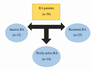 Figure 1. Flowchart of participants in different group |
Results
Totally, 50 RA patients consisted of 5 males and 45 females with an age range of 23 to 65. Table 1 presents the frequency distribution of demographic data, laboratory tests, and clinical parameters across the study groups. The majority of patients were female (n=45, 90%), while 5 (10%) were male. The mean age of female patients was 44.4 ± 10.8 years (Range: 23-65), while that of male patients was 43.4 ± 4.5 years (Range: 39-48). There was no statistically significant difference between groups in terms of gender.
There was no significant difference in demographic characteristics or periodontal parameters among the groups. ESR was the only laboratory parameter that differed significantly among the groups (P-Value=0.016).
The mean hemoglobin level in the Inactive RA group was 12.86 ± 1.85 g/dL (Range: 9.1-16.3 g/dL). In the active RA group, the mean hemoglobin level was 12.95 ± 1.41 g/dL (Range: 10-15 g/dL). The mean hemoglobin level in the recurrent active RA group was 13.16±1.23 g/dl, with a range of 9.9 to 15.3 g/dl. Statistical analysis showed no significant difference in hemoglobin levels among the groups (P-Value=0.724) (Table 1). Based on CAL, 12(24%) patients had mild periodontitis, 25(50%) had moderate periodontitis, and 13(26%) had severe periodontitis.
Furthermore, differences in periodontal parameters between age subgroups were evaluated across all three groups. The Wilcoxon test was used to assess the differences between ESR, CAL, PD, and TL in the age subgroups (Table 2). None of the periodontal indices, including ESR, differed significantly between the age subgroups (≤40 vs. >40 years) across any of the three study groups. It indicates that although the clinical status of RA differed among groups, the periodontal indices did not differ significantly.
|
Table 1. Frequency distribution of demographic data, laboratory tests, and clinical parameters among studied groups (Group 1: Inactive RA; Group 2: Active RA; and Group 3: Recurrent active RA).
 1Bleeding on Probing 2 Clinical Attachment Level 3 Pocket Depth 4Tooth Loss 5 Rheumatoid Factor *Fisher's exact test **Kruskal-Wallis H test |
|
Table 2. Mean rank of periodontal parameters across age subgroups
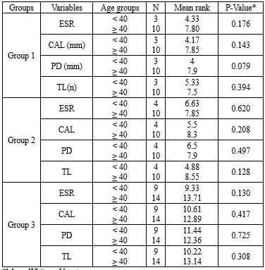 *Mann-Whitney U test |
Discussion
Various studies have emphasized the existence of common genetic factors between RA and periodontitis (10-13,28). In humans, the regulatory genes of the monocytic cytokine response are located in the HLA-DR region of chromosome 5, within the TNF-β area. These genes are implicated in both chronic periodontitis and RA (10).
Previous studies have shown that peptidyl arginine deiminase (PAD) is involved in the pathogenesis of autoimmune damage in RA (29). It is produced by some human cells, such as lung cells, as well as by the microorganism Porphyromonas gingivalis, which supports the association between periodontitis and RA (30-32). The concentration of anti-citrullinated protein antibody (ACPA) and RF is elevated in the serum of patients with RA who are affected by chronic periodontitis (13). Porphyromonas gingivalis generates the ACPA in RA patients, suggesting a direct intersection between periodontitis and RA (33).
Some studies have considered CRP and ESR as components of DAS28 (Disease Activity Score) in their studies to classify RA patients (34,35). Despite this, a large United States registry of RA patients showed that acute phase reactant (APR) levels often do not correlate with disease activity as measured by joint counts and global assessments (36). Furthermore, ESR and CRP are not pathognomonic for RA activity, as they can be evaluated in all inflammatory conditions and infectious conditions (37). Therefore, clinical criteria formed the basis of classification. In 2020, both the European Federation of Periodontology and the American Academy of Rheumatology concluded that there was minimal evidence of a direct association between the two diseases (38).
Periodontitis is more prevalent in individuals in the earliest stages of seropositive RA (39). Kordtabar et al. reported that the gingival indices in RA patients who received anti-rheumatic drugs were significantly lower than in those who did not take medicine. The gingival index in patients with a history of consuming anti-rheumatic drugs was significantly lower than in those who were not drug consumers, showing the effect of the medications on the signs of inflammation (9).
The results of the current study showed no statistically significant difference in BOP, CAL, PD, and TL among different groups of RA patients. According to the analysis, ESR was the only parameter that differed significantly among the groups. The study’s recruitment setting may explain this observation. Since participants were selected exclusively from a rheumatology clinic, their chief complaint was inherently rheumatic disease rather than oral conditions. Patients experiencing oral manifestations secondary to rheumatic disease likely sought treatment at dental centers instead, which may have influenced the prevalence rates observed in this cohort. Smoking and diabetes, as important confounding variables, were excluded from this study to allow a more rigorous investigation of the possible relationship between these two chronic diseases.
The demographic characteristics of our participants were consistent with those of previous study, indicating that RA was more common in females (38). Methodological differences, particularly regarding age stratification, may account for the variations observed between our findings and previous studies. In the present study, the mean age of participants was 44 years. This aligns closely with the typical age of onset of approximately 40 years, when many autoimmune conditions, such as rheumatoid arthritis (RA), typically become clinically apparent. Consequently, 40 years was selected as the cut-off point for stratifying age groups (40). Furthermore, our sample size (N=50) differed from prior studies, which may have influenced statistical power. This study affirmed the findings of a previous study that showed no correlation between RA and periodontitis status (38). This discrepancy may be attributable to differences in ethnic groups and adjustments for confounding variables across different populations (41).
This study found no significant differences in periodontitis indices among RA groups, which is consistent with previous studies (38,39). In contrast, Khantisopon et al. (22) reported a strong relationship among Thai patients. This discrepancy may be attributed to the high prevalence of periodontitis in the general Thai population (22) or to the different classifications of RA patients in this study.
Almashni et al. (2025) concluded that while marginal bone loss and implant survival rates were comparable between RA and non-RA patients, the RA group exhibited a significantly higher plaque index (PI) (42). While our findings showed no significant difference in tooth loss among the study groups, a key distinction lies in the study population. All participants in the current study were RA patients, whereas Almashni et al. examined differences between RA patients and healthy individuals (42). Given the chronic nature of RA, all participants had been exposed to systemic inflammation for an extended duration, which may explain the lack of significant differences in this study between subgroups compared to studies with healthy controls (43). According to the scoping review, there was a close association between bone loss and inflamed joints as well as with CRP and ESR levels. They found a direct relationship between periodontitis and RA (44). The discrepancy between the two studies may be due to different methods of periodontal assessment and the limitation of the sample size, and insufficient statistical power.
Pischon et al. evaluated the association among RA, oral hygiene, and periodontitis in 57 patients with active RA and 57 healthy individuals. The results showed RA patients had more severe periodontitis than the control group (17). Finally, they reported RA patients had significantly higher CAL. They concluded that oral hygiene may only partially account for this relationship. In this study, oral health status was a confounding factor that was not assessed. Dissick et al. investigated the relationship between periodontitis and RA in 69 patients with RA and 35 healthy individuals, assessing their PD and panoramic radiographs (10). They reported that the prevalence of moderate to severe periodontitis in patients with RA was higher than in controls, independent of age, sex, smoking, or diabetes mellitus (10) Our study was consistent with theirs.
Although Kim et al. found no association between chronic periodontitis and rheumatoid arthritis in the Korean population, they concluded that RA was associated with tooth loss (TL) in younger adults (32). While many studies have discussed the relationship between these two diseases and found a significant relationship between CAL, PI, BOP, and PD (45,46). A study found no noteworthy relationship between periodontitis and RA, which is consistent with our findings (1). Wei et al. evaluated a novel target, cathepsin K (Ctsk)-mediated TLR9-related autophagy, during the progression of periodontitis in RA patients (47). Their study utilized a mouse model of periodontitis with RA. Small interfering RNA (siRNA) and CpG oligodeoxynucleotides (CpG ODN) were administered to macrophages. The results showed that RA can promote periodontitis bone destruction in the lesion area. Macrophage stimulation experiments confirmed the in vivo results. This study, the findings of which differ from ours, identified a novel role for Ctsk in TLR9-mediated autophagy to explain the interaction between periodontitis and RA (47).
Rodríguez-Lozano et al. evaluated the association between periodontitis severity and clinical activity in rheumatoid arthritis patients in a case-control study (48). Periodontal exposure was assessed using the following periodontal parameters: PI, BOP, PD, and CAL. They showed a significant association between periodontitis and RA with all periodontal parameters being significantly worse in RA patients than in controls. Periodontitis severity was significantly associated with RA disease activity (48). The inconsistency between their results and ours could be explained by the limited sample size, the wide age range of patients, and the use of different statistical tests.
Future research should assess the diagnosis of periodontitis, the presence of serum ACPA, and their association with the clinical manifestations of RA. The relationship between periodontitis treatment and the reduction of the risk of RA with ACPA positivity should also be examined (13).
Since the data interval was highly dispersed with respect to gender and age range, and the probability of detecting correlations with these variables was low, no significant relationship between different groups was found in the present study. Therefore, future studies with groups matched for periodontal status and with a narrower age range would yield definitive results.
In this study, with a clinical approach, hemoglobin was categorized using established clinical thresholds (WHO criteria for anemia: <12, 12-13, >13 g/dL) to ensure findings are directly interpretable for clinical decision-making. While the authors acknowledge the statistical implications, this approach prioritized clinical utility over continuous analysis in this pilot study.
Although TL in the recurrent active group (23.36%) was higher than in the inactive group (12.83%), there was no statistically significant difference. Due to RA-related disability, such as joint involvement, these patients may have difficulty maintaining good oral hygiene. Dentists play a vital role in achieving and maintaining optimal oral health through timely oral and dental examinations. Despite the many studies conducted on this topic, there is little evidence concerning a possible correlation between RA and periodontitis (39).
Despite its limitations, the study is important and novel in its new approach to stratifying RA patients for the design of future studies. The results showed no statistically significant difference in periodontal indices among the RA groups. Although routine oral health care may seem sufficient for these patients, a specialized protocol for RA patients is needed. This work can be a valuable foundation for the treatment of RA patients.
However, future studies should investigate ACPA and its association with periodontitis. The effect of periodontal treatment on RA parameters and RA duration since diagnosis is another relevant topic for investigation. Additional items, such as periodontal indices, may provide further insight into particular aspects of the disease and facilitate better clinical decision-making with the patient. The absence of formal effect size measures is a limitation of this study, and we strongly recommend their inclusion in future prospective research on this topic. Significant findings, particularly for laboratory parameters, should be interpreted as preliminary and require validation in future, adequately powered studies with pre-specified post-hoc protocols.
The current study must be considered in light of certain limitations. A limitation of this study is the marked gender imbalance, with 90% of the participants being female. While this distribution reflects the higher systemic prevalence of rheumatoid arthritis in women, the findings may not be fully generalizable to male patients, who may exhibit different inflammatory profiles or periodontal progression rates. It is worth mentioning that although a cross-sectional pilot design is appropriate for exploratory analysis, it has limitations regarding causal inference.
The wide age range could be a confounding factor due to the possibility of age-related periodontitis in older patients. Additionally, smoking, BMI, medication type, and disease duration were not adequately controlled and should be addressed in future studies (39).
Conclusion
Our analysis revealed no significant differences in periodontal suggest that among RA patients with active, inactive, or recurrent disease status, whereas ESR levels differed significantly across these groups. This discrepancy indicates that while systemic inflammatory markers such as ESR closely reflect RA disease activity, periodontal parameters may not serve as a reliable surrogate marker for disease flares in this context. Given the cross-sectional nature of this study, these findings should be interpreted as hypothesis-generating rather than definitive evidence of association or causality. Consequently, clinical recommendations implying a probable link between periodontal status and RA activity levels should be made with caution. Future longitudinal studies are required to determine whether periodontal intervention can meaningfully influence RA disease progression.
Acknowledgement
We would like to express our special thanks to Dr. Ali Dehghan, who helped us with useful knowledge and guidance in advancing this study.
Funding sources
This study received no external funding.
Ethical statement
This study was approved by the Ethics Committee of Semnan University of Medical Sciences (Approval code: IR.SEMUMS.REC.1399.193). All enrolled participants were informed about the objectives of this study and signed a written informed consent. All methods were conducted in accordance with the ethical standards of the Declaration of Helsinki.
Conflicts of interest
The authors declare no conflicts of interest.
Author contributions
F.O developed the initial concept of the study. F.O and M.J.S contributed to the study design and wrote the manuscript. M.J.K collected the data, and F.O. oversaw the data collection. S.H.S contributed to the methodology, analysis, and interpretation of data. E.K and F.O contributed to data analysis. All authors revised and approved the final manuscript.
Data availability statement
The data used to support the findings of this study are available from the corresponding author upon request.
Use of artificial intelligence
The authors used an artificial intelligence (AI)-assisted language tool solely to improve the readability, grammar, and linguistic quality of the manuscript. The AI tool was not used to generate or interpret scientific content, design the study, collect or analyze data, or draw conclusions. The authors carefully reviewed and edited the output and take full responsibility for the accuracy, integrity, and originality of the final manuscript.
Editorial: Original article |
Subject:
Health
Received: 2025/11/20 | Accepted: 2025/12/25 | Published: 2025/12/28
Received: 2025/11/20 | Accepted: 2025/12/25 | Published: 2025/12/28
References
1. Dolcezza S, Flores-Fraile J, Lobo-Galindo AB, Montiel-Company JM, Zubizarreta-Macho Á. Relationship Between Rheumatoid Arthritis and Periodontal Disease-Systematic Review and Meta-Analysis. J Clin Med. 2024;14(1):10. [View at Publisher] [DOI] [PMID] [Google Scholar]
2. Jahid M, Khan KU, Rehan Ul H, Ahmed RS. Overview of Rheumatoid Arthritis and Scientific Understanding of the Disease. Mediterranean journal of rheumatology. 2023;34(3):284-91. [View at Publisher] [DOI] [PMID] [Google Scholar]
3. Łasica A, Golec P, Laskus A, Zalewska M, Gędaj M, Popowska M. Periodontitis: etiology, conventional treatments, and emerging bacteriophage and predatory bacteria therapies. Front Microbiol. 2024;15:1469414. [View at Publisher] [DOI] [PMID] [Google Scholar]
4. Huang X, Xie M, Xie Y, Mei F, Lu X, Li X, et al. The roles of osteocytes in alveolar bone destruction in periodontitis. J Transl Med. 2020;18(1):479. [View at Publisher] [DOI] [PMID] [Google Scholar]
5. Ishikawa Y, Terao C. The Impact of Cigarette Smoking on Risk of Rheumatoid Arthritis: A Narrative Review. Cells. 2020;9(2):475. [View at Publisher] [DOI] [PMID] [Google Scholar]
6. Mole EN, Tarassi K, Tsirogianni A, Athanassiades T, Kitsiou V, Kouniaki D, et al. Impact of HLA-DRB1 SE, anti-citrullinated protein antibodies and smoking on radiographic outcome in Greek patients with Rheumatoid Arthritis. BMC Rheumatol. 2025;9(1):128. [View at Publisher] [DOI] [PMID] [Google Scholar]
7. Zhao X, Liu Z, Shu D, Xiong Y, He M, Xu S, et al. Association of Periodontitis with Rheumatoid Arthritis and the Effect of Non-Surgical Periodontal Treatment on Disease Activity in Patients with Rheumatoid Arthritis. Med Sci Monit. 2018;24:5802-10. [View at Publisher] [DOI] [PMID] [Google Scholar]
8. Atarbashi-Moghadam F, Rashidi Maybodi F, Dehghan A, Haerian Ardakani A. Effect of non-surgical periodontal treatment on clinical signs of rheumatoid arthritis. J Adv Periodontol Implant Dent. 2018;10(1):13-7. [View at Publisher] [DOI] [PMID] [Google Scholar]
9. Kordtabar S, Aghaie M, Fakhari E, Vakili MA. Periodontal Condition in Patients with Rheumatoid Arthritis: Effect of Anti-rheumatic Drugs. J Dent (Shiraz). 2019;20(3):190-4. [View at Publisher] [DOI] [PMID] [Google Scholar]
10. Dissick A, Redman RS, Jones M, Rangan BV, Reimold A, Griffiths GR, et al. Association of periodontitis with rheumatoid arthritis: a pilot study. J Periodontol. 2010;81(2):223-30. [View at Publisher] [DOI] [PMID] [Google Scholar]
11. Fathi HM, Khairy N, Gheita TA, El Battawy WA, Taha R. Clinical significance of periodontitis in rheumatoid arthritis patients: association with disease activity and functional status. Journal of Arthritis. 2018;7(2):1-7. [View at Publisher] [DOI] [Google Scholar]
12. Detert J, Pischon N, Burmester GR, Buttgereit F. The association between rheumatoid arthritis and periodontal disease. Arthritis Res Ther. 2010;12(5):218. [View at Publisher] [DOI] [PMID] [Google Scholar]
13. Payne JB, Golub LM, Thiele GM, Mikuls TR. The link between periodontitis and rheumatoid arthritis: a periodontist's perspective. Curr Oral Health Rep. 2015;2(1):20-9. [View at Publisher] [DOI] [PMID] [Google Scholar]
14. Maybodi FR, Bashiri H, Sezavar K, Owlia F. Effect of periodontal treatment on serum inflammatory parameters and disease activity in patients with systemic lupus erythematosus: A randomized controlled trial. J Indian Soc Periodontol. 2022;26(6):564-9. [View at Publisher] [DOI] [PMID] [Google Scholar]
15. Schulz S, Pütz N, Jurianz E, Schaller HG, Reichert S. Are There Any Common Genetic Risk Markers for Rheumatoid Arthritis and Periodontal Diseases? A Case-Control Study. Mediators Inflamm. 2019;2019:2907062. [View at Publisher] [DOI] [PMID] [Google Scholar]
16. Renvert S, Berglund JS, Persson GR, Söderlin MK. The association between rheumatoid arthritis and periodontal disease in a population-based cross-sectional case-control study. BMC Rheumatol. 2020;4(1):31. [View at Publisher] [DOI] [PMID] [Google Scholar]
17. Pischon N, Pischon T, Kröger J, Gülmez E, Kleber BM, Bernimoulin JP, et al. Association among rheumatoid arthritis, oral hygiene, and periodontitis. J Periodontol. 2008;79(6):979-86. [View at Publisher] [DOI] [PMID] [Google Scholar]
18. England BR, Tiong BK, Bergman MJ, Curtis JR, Kazi S, Mikuls TR, et al. 2019 Update of the American College of Rheumatology Recommended Rheumatoid Arthritis Disease Activity Measures. Arthritis Care Res (Hoboken). 2019;71(12):1540-55. [View at Publisher] [DOI] [PMID] [Google Scholar]
19. Smolen JS, Breedveld FC, Burmester GR, Bykerk V, Dougados M, Emery P, et al. Treating rheumatoid arthritis to target: 2014 update of the recommendations of an international task force. Ann Rheum Dis. 2016;75(1):3-15. [View at Publisher] [DOI] [PMID] [Google Scholar]
20. Roodenrijs NMT, Kedves M, Hamar A, Nagy G, van Laar JM, van der Heijde D, et al. Diagnostic issues in difficult-to-treat rheumatoid arthritis: a systematic literature review informing the EULAR recommendations for the management of difficult-to-treat rheumatoid arthritis. RMD Open. 2021;7(1):e001511. [View at Publisher] [DOI] [PMID] [Google Scholar]
21. Cetinkaya B, Guzeldemir E, Ogus E, Bulut S. Proinflammatory and anti‐inflammatory cytokines in gingival crevicular fluid and serum of patients with rheumatoid arthritis and patients with chronic periodontitis. J Periodontol. 2013;84(1):84-93. [View at Publisher] [DOI] [PMID] [Google Scholar]
22. Khantisopon N, Louthrenoo W, Kasitanon N, Sivasomboon C, Wangkaew S, Sang‐In S, et al. Periodontal disease in Thai patients with rheumatoid arthritis. Int J Rheum Dis. 2014;17(5):511-8. [View at Publisher] [DOI] [PMID] [Google Scholar]
23. Al-Jabi SW, Seleit DI, Badran A, Koni A, Zyoud SH. Impact of socio-demographic and clinical characteristics on functional disability and health-related quality of life in patients with rheumatoid arthritis: a cross-sectional study from Palestine. Health Qual Life Outcomes. 2021;19(1):241. [View at Publisher] [DOI] [PMID] [Google Scholar]
24. Shakeel L, Shaukat A, Khaliq N, Kashif A, Mujeeb A, Adnan Z, et al. Rheumatoid arthritis: a comprehensive overview of genetic markers, emerging therapies, and personalized medicine. Ann Med Surg (Lond). 2025;87(2):696-710. [View at Publisher] [DOI] [PMID] [Google Scholar]
25. Mohammadi.B.A A, Karami M, Nouri M, Morovati SP. Comparison of the efficiency of three tooth brushing training methods on dental plaque removal of 10-year-old female students. ijpd. 2022;17(1):1-11. [View at Publisher] [Google Scholar]
26. Cheng X, Liu J, Liu S, Fang D, Chen X, Ding X, et al. Red Blood Cell-Related Parameters in Rheumatoid Arthritis: Clinical Value and Immunological Significance. J Inflamm Res. 2024;17:10641-50. [View at Publisher] [DOI] [PMID] [Google Scholar]
27. Bosma ML, McGuire JA, DelSasso A, Milleman J, Milleman K. Efficacy of flossing and mouth rinsing regimens on plaque and gingivitis: a randomized clinical trial. BMC Oral Health. 2024;24(1):178. [View at Publisher] [DOI] [PMID] [Google Scholar]
28. O'Leary TJ, Drake RB, Naylor JE. The plaque control record. J Periodontol. 1972;43(1):38-. [View at Publisher] [DOI] [PMID] [Google Scholar]
29. Reyes-Castillo Z, Muñoz-Valle JF, Llamas-Covarrubias MA. Clinical and immunological aspects of anti-peptidylarginine deiminase type 4 (anti-PAD4) autoantibodies in rheumatoid arthritis. Autoimmun Rev. 2018;17(2):94-102. [View at Publisher] [DOI] [PMID] [Google Scholar]
30. Koziel J, Mydel P, Potempa J. The link between periodontal disease and rheumatoid arthritis: an updated review. Curr Rheumatol Rep. 2014;16(3):408. [View at Publisher] [DOI] [PMID] [Google Scholar]
31. Fuggle NR, Smith TO, Kaul A, Sofat N. Hand to mouth: a systematic review and meta-analysis of the association between rheumatoid arthritis and periodontitis. Front Immunol. 2016;7:80. [View at Publisher] [DOI] [PMID] [Google Scholar]
32. Kim J-W, Park J-B, Yim HW, Lee J, Kwok S-K, Ju JH, et al. Rheumatoid arthritis is associated with early tooth loss: results from Korea National Health and Nutrition Examination Survey V to VI. Korean J Intern Med. 2019;34(6):1381-91. [View at Publisher] [DOI] [PMID] [Google Scholar]
33. de Molon RS, Rossa Jr C, Thurlings RM, Cirelli JA, Koenders MI. Linkage of periodontitis and rheumatoid arthritis: current evidence and potential biological interactions. Int J Mol Sci. 2019;20(18):4541. [View at Publisher] [DOI] [PMID] [Google Scholar]
34. Nasir N, Majid H, Khan AH, Awan S, Riaz MM. Disease activity correlates and functionality in patients with rheumatoid arthritis - real-world experience from a South Asian country. Reumatologia. 2022;60(3):183-91. [View at Publisher] [DOI] [PMID] [Google Scholar]
35. Zhang J, Wang Y, Nie W, Li Q, Li S-G, Jin D. CBC‑derived inflammatory indices for rheumatoid arthritis diagnosis and activity assessment: differential performance by serostatus. Front Immunol. 2026;17:1740898. [View at Publisher] [DOI] [PMID] [Google Scholar]
36. Almazroea AH, Yousef S, Ahmad SM, AlHiraky HN, Al-Haidose A, Abdallah AM. The Impact of ACE Gene Variants on Acute-Phase Reactants in Children with Rheumatic Heart Disease. Diagnostics. 2023;13(10):1672. [View at Publisher] [DOI] [PMID] [Google Scholar]
37. Leong KP, Tan JWL, Gao X, Koh ET. Conversion among the 28-joint count activity indices for rheumatoid arthritis. Eur J Rheumatol . 2020;7(3):105-11. [View at Publisher] [DOI] [PMID] [Google Scholar]
38. Bartold PM, Lopez‐Oliva I. Periodontitis and rheumatoid arthritis: an update 2012‐2017. Periodontol 2000. 2020;83(1):189-212. [View at Publisher] [DOI] [PMID] [Google Scholar]
39. Ceccarelli F, Saccucci M, Di Carlo G, Lucchetti R, Pilloni A, Pranno N, et al. Periodontitis and rheumatoid arthritis: the same inflammatory mediators? Mediators Inflamm. 2019:2019:6034546. [View at Publisher] [DOI] [PMID] [Google Scholar]
40. Pawłowska J, Smoleńska Z, Daca A, Witkowski JM, Bryl E. Older age of rheumatoid arthritis onset is associated with higher activation status of peripheral blood CD4(+) T cells and disease activity. Clin Exp Immunol. 2011;163(2):157-64. [View at Publisher] [DOI] [PMID] [Google Scholar]
41. Rovas A, Puriene A, Punceviciene E, Butrimiene I, Stuopelyte K, Jarmalaite S. Associations of periodontal status in periodontitis and rheumatoid arthritis patients. J Periodontal Implant Sci. 2021;51(2):124-34. [View at Publisher] [DOI] [PMID] [Google Scholar]
42. Almashni H, Kakar E, Nava P, Wang HL, Saleh MHA. Influence of rheumatoid arthritis on peri-implant diseases: A longitudinal retrospective clinical and radiographic evaluation. J Periodontol. 2025;96(8):933-43. [View at Publisher] [DOI] [PMID] [Google Scholar]
43. Auyezkhankyzy D, Izekenova A, Kocyigit BF. Emergencies in inflammatory rheumatic diseases. Rheumatol Int. 2024;44(9):1637-46. [View at Publisher] [DOI] [PMID] [Google Scholar]
44. Nik-Azis N-M, Mohd N, Baharin B, Said MSM, Fadzilah FM, Haflah NHM. Periodontal Disease in Seropositive Rheumatoid Arthritis: Scoping Review of the Epidemiological Evidence. Germs. 2021;11(2):266-86. [View at Publisher] [DOI] [PMID] [Google Scholar]
45. Angiolani F, Gerardi D, Bernardi S, Torge D, Bianchi S, Piattelli M, et al. Bidirectional Relationship Between Periodontal Disease and Reproductive Disorders: Focus on Polycystic Ovary Syndrome. Oral. 2025;5(3):67. [View at Publisher] [DOI] [Google Scholar]
46. Ceraulo S, Barbarisi A, Caccianiga G, Lodico M, Cremonini F, Lauritano D, et al. A possible correlation between periodontal disease and systemic diseases: a clinical study. BMC Oral Health. 2025;26(1):13. [View at Publisher] [DOI] [PMID] [Google Scholar]
47. Wei W, Ren J, Yin W, Ding H, Lu Q, Tan L, et al. Inhibition of Ctsk modulates periodontitis with arthritis via downregulation of TLR9 and autophagy. Cell Prolif. 2020;53(1):e12722. [View at Publisher] [DOI] [PMID] [Google Scholar]
48. Rodríguez-Lozano B, González-Febles J, Garnier-Rodríguez JL, Dadlani S, Bustabad-Reyes S, Sanz M, et al. Association between severity of periodontitis and clinical activity in rheumatoid arthritis patients: a case-control study. Arthritis Res Ther. 2019;21(1):27 [View at Publisher] [DOI] [PMID] [Google Scholar]
Send email to the article author
| Rights and permissions | |
 |
This work is licensed under a Creative Commons Attribution-NonCommercial 4.0 International License. |